PHONE
+44-7482-878921
+44-7482-878921
2376-0249
Clinical-Medical Image - International Journal of Clinical & Medical Images (2020) Volume 7, Issue 3

Author(s): Jan Jonckheere*, Hendrik Everaert, Dirk Van Den Berge, Lode Goethals, Seema Doring, Johan de Mey
NF type 3 is a rare form of neurofibromatosis.
If multiple hypermetabolic lesions are present on PET/CT, like schwannomas in NF type 3, benign pathology should always be considered and should not be mistaken for malignant metastatic disease.
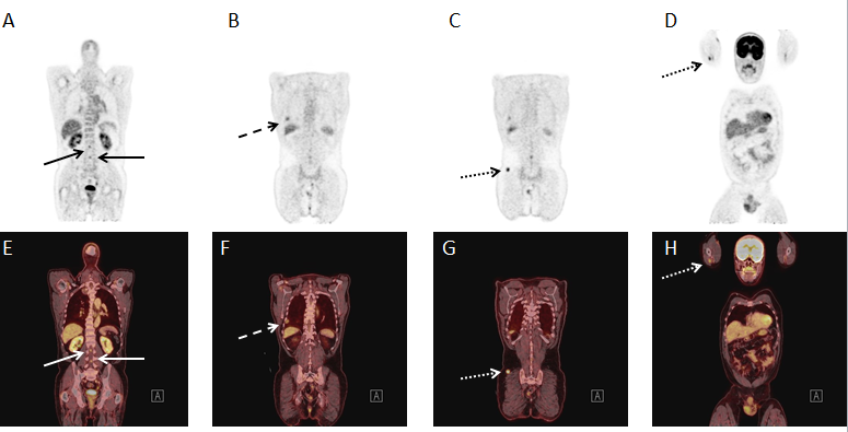
PET/CT was performed in a 56-year-old male patient to exclude metastatic disease. Coronal views on PET (panels A, B, C and D) and on integrated PET/CT (panels E, F, G and H) demonstrate increased F-18 FDG uptake in lesions located in the spinal canal at lumbar level (normal arrow), in the right pleura (dashed arrow), in the biceps muscle of the right arm (dotted arrow) and in the right gluteus muscle (dotted arrow) with a distribution pattern along the peripheral nerves (Figure 1).
PET/CT is very useful in planning a biopsy to confirm the diagnosis because of a good overview of all lesions so the safest lesion to biopsy can be determined. The distribution pattern of different rounded, sharply demarcated lesions along the peripheral nerves, suggests benign pathology with schwannomas. Schwannomas can show a wide variation of SUV values so these values are less contributive in differentiation from metastatic disease.
Additional CT-guided biopsy confirmed the diagnosis of NF type 3.
 Awards Nomination