PHONE
+44-7482-878921
+44-7482-878921
2376-0249
Case Blog - International Journal of Clinical & Medical Images (2015) Volume 2, Issue 9

Author(s): Chih-Cheng Lu and Chien-Ming Chao*
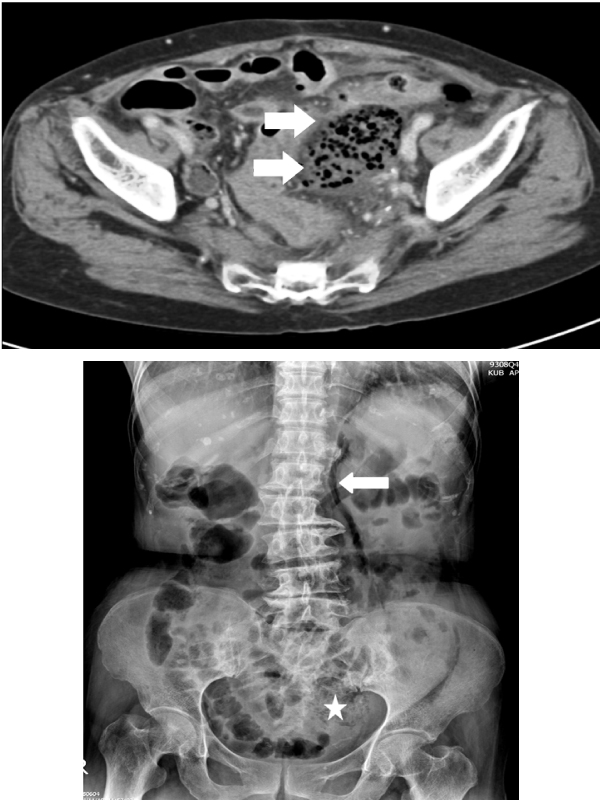
Case: A 58 year-old woman presented with fever, and right flank pain for two days. In addition, gross hematuria was noted this morning. The patient had a history of diabetes mellitus. On arrival, her vital signs were temperature of 39°C, pulse rate of 127/min, respiratory rate of 24/min, and blood pressure of 96/65 mm Hg. Physical examinations only disclosed mild right flank knocking tenderness. Laboratory examinations were as follows: white cell count 22,300/mm3 with predominance of neutrophils (97%), platelet 30,000/mm3 , C-reactive protein of 269 mg/L (reference value, less than 3 mg/L). Urinary analysis showed both of white blood cells and red blood cells greater than 100/high power field. Radiography of the abdomen revealed air within right ureter and in the right kidney area (Figure 1).
Computed tomography of the abdomen disclosed emphysematous pyelonephritis with concomitant emphysematous ureteritis and a ureteral stone (Figure 2). Therefore, the patient received antibiotic with flomoxef for severe urosepsis and emergent placement of a percutaneous nephrostomy. Three days later, all of her blood cultures, and pus collected from the drain grew Escherichia coli. Thereafter, the clinical condition became stable and she was discharged two weeks later. Emphysematous urinary tract infection is a necrotizing infection, which is characterized by the accumulations of air in the renal parenchyma, collecting system or urinary bladder.
E. coli and Klebisella pneumoniae are the most common pathogens, and diabetes mellitus is major risk factor. The mortality may be up to 18-40%, and prompt management including systemic antibiotics and immediate drainage or surgical intervention, are essential for life-saving [1]. Image study such as a simple KUB or CT can be helpful for diagnosis.
 Awards Nomination