PHONE
+44-7482-878921
+44-7482-878921
2376-0249
Case Blog - International Journal of Clinical & Medical Images (2015) Volume 2, Issue 3

Author(s): Alexander KC Leung* and Benjamin Barankin
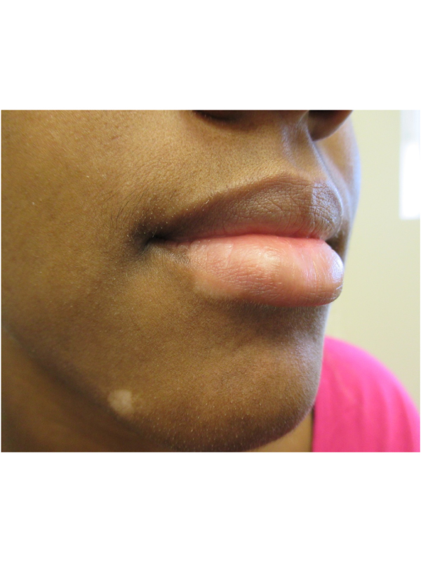
Case: A 21-year-old man presented with white macules and patches on his lower lip and chin for the past 5 years. The lesions were asymptomatic but cosmetically unsightly. His health was otherwise unremarkable. There was no family history of a similar skin disorder or autoimmune disease. Vitiligo is a common acquired pigmentation disorder characterized by depigmented macules and patches as a result of loss of functional cutaneous melanocytes. The condition affects 0.5 to 1% of the population worldwide. Approximately 25% of affected patients have the onset of vitiligo before the age of 10 years and 50% before the age of 20 years. Genetic, immunological, and neurogenic factors may act alone or synergistically to trigger or perpetuate the loss of functional melanocytes from the basal layer of the epidermis. Typically, vitiligo presents as acquired amelanotic macules and patches that appear chalk or milk-white in color. Lesions often show homogenous depigmentation and are well demarcated. Lesions are often symmetrical and enlarge centrifugally in size with time.
The most common location is the face, followed by the neck, lower limbs, trunk, and upper limbs. The diagnosis is mainly clinical, based on the findings of acquired, well-demarcated white macules and patches that tend to enlarge. Wood’s lamp accentuates the lesion and may be of benefit in the diagnosis (e.g. to differentiate from tinea versicolor or pityriasis alba), especially in individuals with skin type I and type II. The clinical course is generally unpredictable. In individuals with skin type I and type II, no active treatment other than the use of camouflage cosmetics and sunscreens is usually recommended. Psychological support should be offered when necessary. If treatment is preferred for cosmetic reasons, a variety of treatment options are available. These include topical corticosteroids, topical calcineurin inhibitors, narrowband ultraviolet B phototherapy (nbUVB), and monochromatic excimer laser and light at 308 nm.
 Awards Nomination