PHONE
+44-7482-878921
+44-7482-878921
2376-0249
Case Blog - International Journal of Clinical & Medical Images (2018) Volume 5, Issue 11

Author(s): Tricarico Laura*
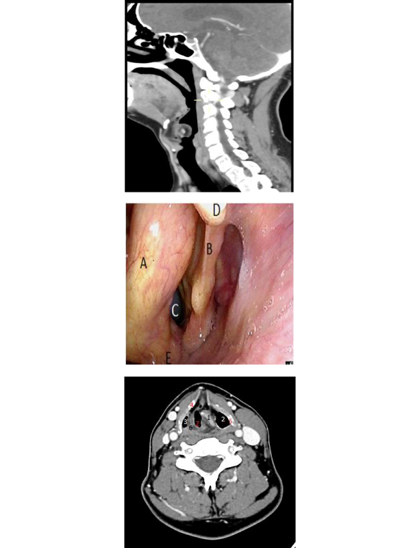
Clinical Image: A 45-year-old male subject with recent diagnosis of right colon cancer was referred to Department of Otorhinolaryngology for a preoperative airway assessment. The patient presented a long years history of intermittent hoarseness, retrosternal burning and rare episodes of laryngospasm, which however have never been further investigated. Videolaryngoscopy examination (Figure 1) showed a pinkish, smooth and hard mass in the supraglottis, covered by regular mucosa, narrowing anterior glottic region, which demonstrated the left vocal fold fixed in paramedial position. The subglottic space appeared well preserved. CT scan of the neck (Figures 2 and 3) revealed an hypodense mass originating from left aryepiglottic fold incontiguity with the left arytenoids and expanded from the posteromedial left side to superomedial side. The lesion presented a peripheral calcified stippled appearance and was surrounded by discreet soft -tissue masses, without enhancement of intravenous contrast. No enlarged lymph nodes were visible. The imaging findigs are indicative of a congenital laryngeal malformation.The airway space was judged sufficient for endotracheal intubation to perform abdominal surgery. No introperative or extubation complications occured. The patient will be introduced to a dedicated follow-up endoscopy to carry out biopsy and surgical treatment of the laryngeal lesion.
 Awards Nomination