PHONE
+44-7482-878921
+44-7482-878921
2376-0249
Clinical-Medical Image - International Journal of Clinical & Medical Images (2022) Volume 9, Issue 6

Author(s): Shanthi Mariapan*, Sangaran Gopal, Ruchi Negi, Fatin Haziqah and Dinie Tumaisuri
Hospital Shah Alam, Shah Alam, Selangor, Malaysia
Received: 06 June, 2022, Manuscript No. ijcmi-22-21740; Editor assigned: 08 June, 2022, 2022, PreQC No. P-21740; Reviewed: 16 June, 2022, QC No. Q-21740; Revised: 23 June, 2022, Manuscript No. R-21740; Published: 30 June, 2022, DOI: 10.4172/2376-0249.1000832
Citation: Mariapan S, Sangaran G, Negi R, Haziqah F and Tumaisuri D. (2022) A Case of External Ear and Submandibular Extracranial AVM. Int J Clin Med Imaging 9:832.
Copyright: © 2022 Mariapan S, et al. This is an open-access article distributed under the terms of the Creative Commons Attribution License, which permits unrestricted use, distribution, and reproduction in any medium, provided the original author and source are credited.
Introduction
Vascular anomalies are classified as vascular tumor such as hemangioma and vascular malformation. Vascular malformation is of low-flow and high-flow and may arise from blood vessels or lymph vessels. Vascular tumors are true neoplasms with abnormal cellular turnover. Vascular malformations on the other hand, do not undergo pathologic cell proliferation. Arteriovenous malformation of the head and neck is a rare vascular anomaly but when present is persistent and progressive in nature due to failure of regression of primitive arteriovenous channels during 4th till 6th week gestation.
Objective
The aim of this case report is to show the progressiveness of an extracranial AVM of a patient over the course of 20 years.
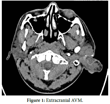
Encik F, 34 years old gentleman, first presented with a swelling at the left ear pinna since the age of 15 years old (pubertal age) in the year 2000. In 2006, he noted dilated vessels traversing this swelling. Another swelling at the left cheek was also noted at this time frame. No aggravating factor. No history of trauma therefore it was not a hematoma. Not associated with pain. He had defaulted treatment in HKL since 2012 (initially planned for embolisation and total pinectomy) due to missed appointment. A set of investigation was done at that time and he was diagnosed as extracranial AVM. On 6.8.2019 there was sudden blood spurting from left pinna and he rushed back to Hospital Shah Alam where the bleeding was arrested by compression. No headache. No shortness of breath. Upon further history, his swelling has increased in size from 2012. It doesn’t affect day to day activity. No other past medical illness or surgical history. On examination in August 2019 noted that the whole left pinna was pulsating with post auricular, infra auricular and left submandibular pulsating mass measuring 2 ×3 cm. It was soft, colourless and not warm to touch. His platelet in August 2019 was 197, WBC was 6.8. Further investigation in August showed an INR of 0.98, and there is no prolonged PT or APTT at that time to suggest clotting abnormalities. Upon CT angiography Carotid, noted that the Facial branch, Maxillary branch, Posterior Auricular and Superficial Temporal branch of the left External Carotid artery appear dilated and tortuous. There is abnormal clump of dilated vessels (arteriovenous malformation) at the level of left submandibular and left external ear. The feeding arteries are from the left Facial artery for the left submandibular AVM and left Maxillary as well as Superficial Temporal arteries for left external ear AVM. The draining veins are identified as the dilated left External Jugular vein as well its branches at the left submandibular and left external ear/ peri auricular region. The left external ear is swollen. Minimal punctate calcification seen at the left ear pinna. He was referred back to hkl- Hospital Kuala Lumpur for treatment. Patient claimed he was not comfortable with surgical treatment and opted out of it. However, he noted that there are new dilated vessels behind his ear that is pulsating and increasing in size since early September 2020. He was advised to consult Hospital Kuala Lumpur Plastic surgery department.
53 individual cases of external ear AVM has been reported by various literatures. From these, it is found that the average year of AVM is 26 years old. 7 patients reported that their AVM became aggravated during puberty and 6 had histories of trauma. Meanwhile, according to Raymond SB, et al. [1], the exact incidence of submandibular AVMs is not known but based on case series is estimated at 1 in 50000. Kohout MP, et al. [2] in their study also found that AVMs were present at birth in 59% of cases, in childhood 10% of cases, in adolescent 10% of cases, and in adulthood 21% of cases. In this case, the onset of the AVM was at puberty and progressed from the external ear to the submandibular level involving the external carotid artery branches. Follow up for this patient may provide useful in monitoring further vessel dilatation or new AVM cranially or caudally from the branches of the external carotid artery with the external jugular vein. There was no internal carotid artery involvement or perfusion deficit, no extension onto the upper eyelid, and no obvious atrophy or compression from the AVM onto the submandibular gland [3] (Figure 1).
Extracranial AVM itself is a rare entity. Author was unable to find reported incidence of external ear and submandibular AVM in the same individual, however there was a reported case of infra temporal and parotid AVM. Currently there are more protruding vessels seen at the back of patient’s ear and he was advised to revisit Hospital Kuala Lumpur for treatment option. Therefore, it is a sign of persistence and progressiveness of his disease.
AVM, Extra cranial.
Google Scholar, Crossref, Indexed at
 Awards Nomination