PHONE
+44-7482-878921
+44-7482-878921
2376-0249
Case Blog - International Journal of Clinical & Medical Images (2014) Volume 1, Issue 5

Author(s): Paulos Yigazu
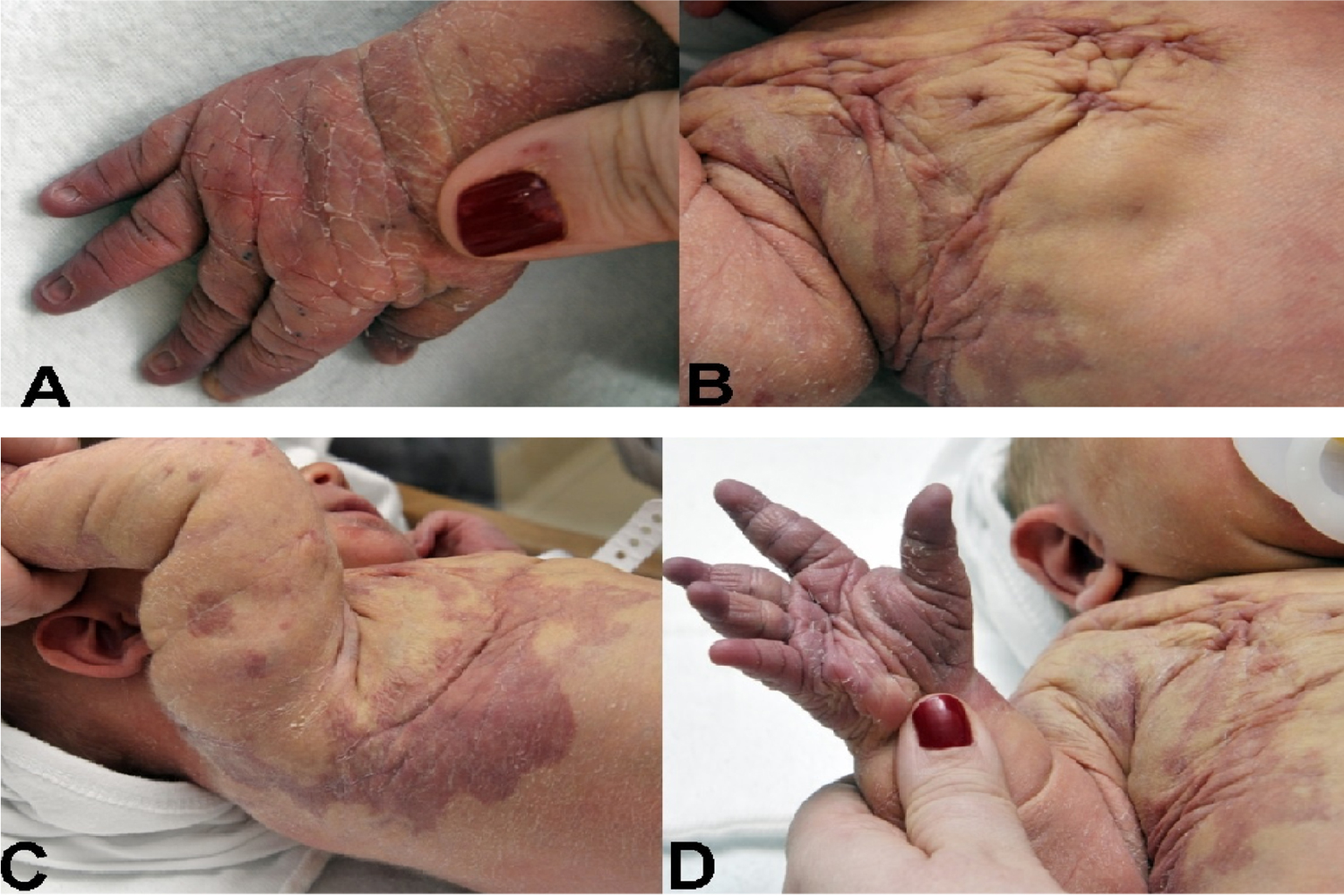
This full-term boy was born to a 41 year old mother by elective Caesarean section. Prenatal ultrasound showed an echo-lucent mass on the right side of the body consistent with possible vascular malformation. Newborn physical examination revealed tissue hypertrophy and desquamation of skin on right hand (Figure 1). In addition to several small hemangiomas noted on scalp and extremities, there was a large hemangiomatous lesion extending from posterior right shoulder to right lateral chest wall below right arm (Figure 2). Ultrasound of the lesion demonstrated multiple cystic areas with internal septations in the soft tissues of the right anterior chest wall extending to the right flank, posterior upper back and right shoulder, and right upper extremity extending to the right elbow. There was no internal flow on color Doppler in these cystic areas. Thickening of the adjacent subcutaneous tissues was noted. These findings were consistent with a lymphatic malformation. Bilateral duplex imaging of the deep and superficial veins of the upper extremities showed no evidence of acute or chronic deep or superficial venous thrombosis. Based on the findings of lymphatic vascular malformation, superficial hemangiomas and tissue hypertrophy, a clinical diagnosis of Klippel-Trenaunay Syndrome was made. Klippel-Trenaunay Syndrome (KTS) is a condition defined by the association of capillary malformation (98%), varicosities or vein malformations (72%), and hypertrophy of bony and soft tissues (67%) [1]. The presence of any two of these three featuresis considered sufficient for diagnosis of KTS [2]. Lymphatic malformations are also a common feature of KTS [2]. Figure Tissue hypertrophy and desquamation of the skin on right hand. Figure A large hemangiomatous lesion extending from posterior right shoulder to right lateral chest wall below right arm.
*Corresponding author: Paulos Yigazu, The Carman and Ann
Adams Department of Pediatrics, Division of Neonatal-Perinatal
Medicine, Wayne State University School of Medicine, Children’s
Hospital of Michigan & Hutzel Women’s Hospital, Detroit, MI 48201,
USA, Tel: (646)-715-8044; E-Mail: paulosnigat2003@yahoo.com
 Awards Nomination