2376-0249
Case Blog - International Journal of Clinical & Medical Images (2016) Volume 3, Issue 2

Author(s): Sushant Soren, Ajay Manickam, Jr Das, Jayanta Saha and Sk Basu
Introduction Papillary thyroid carcinomas are one of the most common endocrine neoplasms. It is well known for its low malignant potential. Papillary carcinomas mostly metastases to lymph nodes of level six. Recent advances in the pathology that is with proper ultra sound guided FNAC, early diagnosis can be made. Here we are presenting a patient with a huge neck mass that presented to our medical college hospital and how we managed the same.
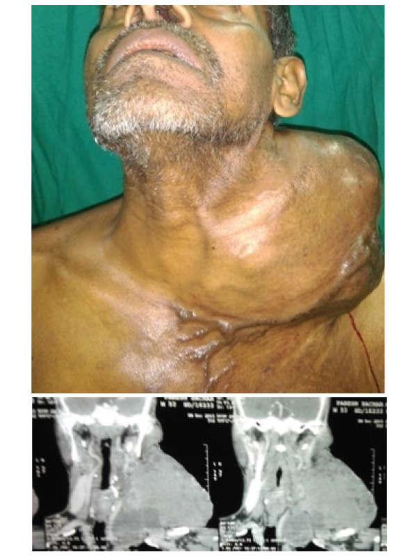
Case Blog 53 years old male patient, presented to a tertiary hospital with complaints of swelling in front of neck for the past 7 years. The patient was having a progressive swelling, it started as a small pea shaped swelling and it slowly progressed and has reached the present size (Figure 1). The patient was having no difficulty in respiration. CT scan of the neck region with chest was taken as the lower extent was not palpable and the FNAC of the swelling was taken. FNAC revealed papillary thyroid carcinoma.
CT scan showed a huge soft tissue mass over left supra clavicular region with solid and cystic attenuation extending to the superior aspect of the thoracic inlet. The mass was shifting trachea to the right side (Figure 2). There were no palpable neck nodes. The patient was planned for surgical exploration of the tumour with sternotomy as the tumour was extending in to the thoracic inlet. A total thyroidectomy with central compartment neck dissection was done. The mass was proved to be papillary thyroid carcinoma of intra cystic variant.
Discussion Papillary thyroid carcinoma is the most common thyroid cancer. Papillary thyroid cancer also has an excellent prognosis. Total thyroidectomy is done once histo pathological diagnosis is established [1].
This patient to begin with presented with a small pea shaped swelling on front of left side of neck and it started slowly progressing in size. Once the size of the tumour is more than 10 mm, total thyroidectomy is indicated. These papillary carcinomas mostly metastases to central neck nodes [2]. Hence for a better outcome it is always mandatory to go for central neck node dissection for proven cases of lymphatic spread. Biochemical analysis of free T4 and TSH is necessary before surgery. This patient was euthyroid before surgery was planned. Surgery of total thyroidectomy and neck dissection of central compartment was done [3]. Recurrent laryngeal nerve was identified during the procedure. Parathyroid glads were identified and preserved on the right side. The patient was kept on regular followup. Histopathological diagnosis confirmed it to be a papillary thyroid carcinoma.
Conclusion Total thyroidectomy with central compartment dissection appears to be adequate treatment even for masses that are extending upto supra clavicle also with extension to thoracic inlet. CT scan plays a very important role in pre-operative planning for huge neck mass. Preservation of recurrent laryngeal nerve and parathyroid glands are very important in this surgery. Though it is controversial in the absence of neck node one should go for central node dissection, from our institutional experience, it is always safer to go for central node dissection, so that recurrence of the tumour can be avoided.
Haberler
Haberler
Haberler
Haberler
Haberler
Haberler
Haberler
Haberler
Haberler
Haberler
Haberler
Haberler
Haberler
Haberler
Haberler
Haberler
Haberler
Haberler
Haberler
Haberler
Haberler
Haberler
Haberler
Haberler
Haberler
Haberler
Haberler
Haberler
Haberler
Haberler
Haberler
Haberler
Haberler
Haberler
Haberler
Haberler
Haberler
Haberler
Haberler
Haberler
Haberler
Haberler
Haberler
Haberler
Haberler
Haberler
Haberler
Haberler
Haberler
Haberler
 Awards Nomination

