PHONE
+44-7482-878921
+44-7482-878921
2376-0249
Medical Image - International Journal of Clinical & Medical Images (2015) Volume 2, Issue 3

Author(s): Wang YW and Lin WT*
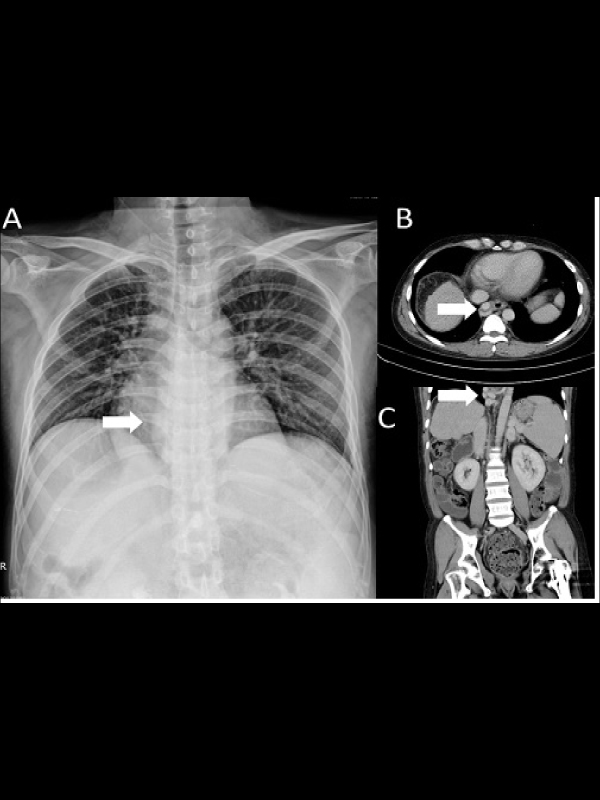
Medical Image: A 33-year-old man presented with massive bloody vomitus. He had the history of alcoholic liver cirrhosis. Emergent esophagogastroenteroscopy found the presence of esophageal varices (EV) with active bleeding and EV ligation was performed. In the meanwhile, outline chest radiography showed a retrocardiac opacity (Figure 1A, arrow). Computed tomography of chest disclosed contrast-enhancing paraesophageal veins just to the left of the esophagus, which was compatible with EV (Figure 1B, and 1C, arrows).
 Awards Nomination