PHONE
+44-7482-878921
+44-7482-878921
2376-0249
Clinical-Medical Image - International Journal of Clinical & Medical Images (2024) Volume 11, Issue 2

Author(s): Eric Michel Charlemagne Junior, Kessi*, Imrani Kaoutar, Kaoutar Maslouhi, Basma Dghoughi, Tsioukaka Gray Delors, Moatassim Billah Nabil and Ittimade Nassar*
Department of Central Radiology, UHC Ibn Sina Mohammed V University, Rabat, Morocco
*Corresponding Author:
Eric Michel Charlemagne Junior Kessi
Department of Central Radiology, UHC Ibn Sina Mohammed V University, Rabat, Morocco
Tel: 212600714720
E-mail: kessieric1@gmail.com, ericmichelkessi@gamil.com
Received: 01 February 2024, Manuscript No. ijcmi-24-126534; Editor assigned: 03 February 2024, Pre QC No. P-126534; Reviewed: 15 February 2024, QC No. Q-126534; Revised: 21 February 2024, Manuscript No. R-126534; Published: 29 February 2024, DOI:10.4172/2376-0249.1000941
Citation: Kessi EMCJ, Imrani K, Maslouhi K, Dghoughi B and Tsioukaka GD, et al. (2024) Anatomical Variant at Surgical Risk: Median Arcuate Ligament
Syndrome. Int J Clin Med Imaging 11: 941.
Copyright: © Kessi EMCJ, et al. This is an open-access article distributed under the terms of the Creative Commons Attribution License, which permits
unrestricted use, distribution, and reproduction in any medium, provided the original author and source are credited.
Median Arcuate Ligament Syndrome (MALS), also known as Dunbar’s syndrome, is a rare condition caused by compression of the Celiac Trunk (CT) by the fibrous attachments of the median arcuate ligament and the diaphragmatic crest. It is an anatomical variant that affects about 10-24% of the population and about 1% develop symptoms [1].
This syndrome was first described in the series by Harjola in 1963 and by Dumbar, et al. in 1965, and thus became known as Dumbar’s syndrome. It is clinically identified by a symptomatic triad associating post-prandial epigastric pain, unexplained weight loss and auscultatory noises (inconstant) following arterial stenosis [2,3]; These pains may or may not be accompanied by nausea, vomiting and diarrhea. However, a small proportion of the general population has a relatively compressive medial arcuate ligament, with or without specific symptoms [4,5]. This asymptomatic nature is often the cause of misdiagnosis, and it often takes a long time before a diagnosis can be made, which is not always obvious.
Our patient had no digestive symptoms associated with significant compression of the celiac trunk. He presented with cholestatic jaundice in a type 2 diabetic patient with general deterioration of condition, requiring abdominal ultrasound, which revealed a mass in the head of the pancreas with dilatation of the bile ducts; further investigations revealed a cervico-isthmic pancreatic tumour process.
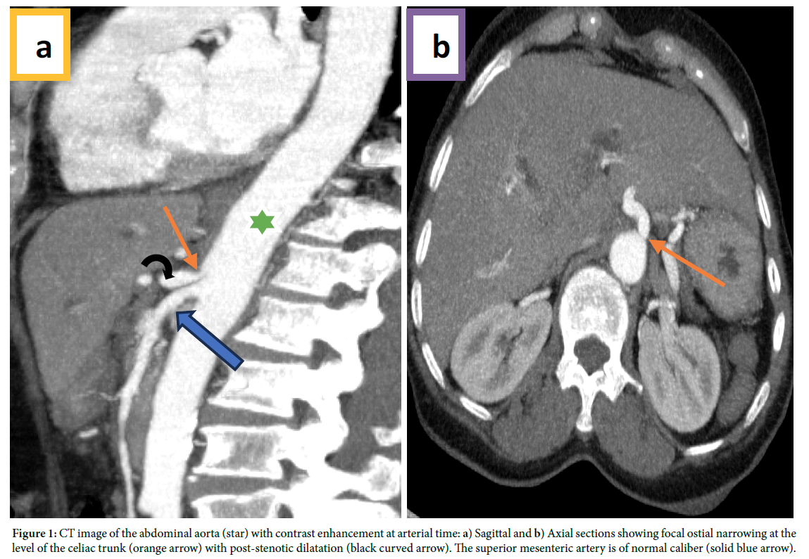
Abdominal angioscanner facilitates diagnosis by demonstrating narrowing of the celiac trunk with post-stenotic dilatation (Figure 1); it allows three-dimensional visualization of the compressed celiac artery; this three-dimensional (3D) shape makes it possible to appreciate the degree of angulation of the CT in relation to the abdominal aorta [1].
This condition is managed surgically, with decompression of the constriction of the medial arcuate ligament of the celiac artery. This can range from ligament transection to aorto-celiac bypass [4].
The frequency between SLAM and pancreatic tumor is high, and resection of a cephalic pancreatic tumor involves resection of the gastroduodenal artery and pancreaticoduodenal arches. However, the latter constitute a bypass between the territories of the CT and the Superior Mesenteric Artery (SMA). In the case of tight stenosis of the CT or SMA, cephalic duodenopancreatectomy is likely to result in supramesocolic arterial ischemia. For this reason, the search for MALS should be systematic and should be mentioned and complemented by a meticulous vascular study, enabling the surgeon to avoid a moribund operation [6].
Median arcuate ligament; Celiac trunk; Compression; Variant; Pancreatic surgery
No conflict of interest.
[1] Abechri A, and Mauel E. Syndrome du ligament arqué médian (de Dunbar): Revue de la littérature sur une errance diagnostique des douleurs abdominales chroniques.
[2] Goodall R, Langridge B, Onida S, Ellis M. and Lane T, et al. (2020) Median arcuate ligament syndrome. J Vasc Surg 71(6): 2170-2176.
Google Scholar, Crossref, Indexed at
[3] Iqbal S, and Chaudhary M. (2021) Median arcuate ligament syndrome (Dunbar syndrome). Cardiovascular Diagnosis and Therapy 11(5): 1172.
Google Scholar, Crossref, Indexed at
[4] Amouzou EG, Sogan A, Geraldo R, Plante R and Gueouguede E, et al. (2021) Syndrome du ligament arqué médian: Une hantise pour le chirurgien hépato-bilio-pancréatique. Journal afr chir digest; 21 (2): 3576-3579
[5] Duffy AJ, Panait L., Eisenberg D, Bell RL. and Roberts KE, et al. (2009) Management of median arcuate ligament syndrome: A new paradigm. Ann Vasc Surg 23(6): 778-784.
Google Scholar, Crossref, Indexed at
[6] Pinto A, Weyl, A, and Carrere N. (2016) Anatomical variant: Median arcuate ligament syndrome, and its consequences on the resectability of head pancreatic tumors. Morphology 100(330): 142.
 Awards Nomination