PHONE
+44-7482-878921
+44-7482-878921
2376-0249
Clinical-Medical Image - International Journal of Clinical & Medical Images (2023) Volume 10, Issue 7

Author(s): Ghita EL Adioui*
Department of Radiology, HER Children’s Hospital of Rabat, CHU IBN SINA, Rabat, Morocco
Received: 14 June 2023, Manuscript No. ijcmi-23-103120; Editor assigned: 15 June 2023, Pre QC No. P-103120; Reviewed: 04 July 2023, QC No. Q-103120; Revised: 10 July 2023, Manuscript No. R-103120; Published: 17 July 2023, DOI:10.4172/2376-0249.1000902
Citation: Adioui GEL. (2023) Antenatal Urinoma Revealing a Posterior Urethral Valve. Int J Clin Med Imaging 10: 902.
Copyright: © 2023 Adioui GEL. This is an open-access article distributed under the terms of the Creative Commons Attribution License, which permits unrestricted use, distribution and reproduction in any medium, provided the original author and source are credited.
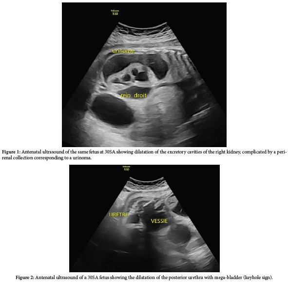
The posterior urethral valve is the most common congenital obstructive lesion of the urethra, with an estimated incidence of ~1 in 10,000- 25,000 live births [1]. Diagnosis can be made antenatally, It is rarely detected in the first trimester, as the prognosis is poor, but more often after 18SA. The urinoma is the ultimate complication and results from the hyper-pressure in the excretory cavities that progress to UHN and rupture of a calyx, resulting in antenatal urinoma (Figure 1), with a poor prognosis. In addition, the distension of the posterior urethra and a megabladder (Figure 2) is very useful for confirming the etiological diagnosis of this sub vesical obstruction [2]. Other suggestive signs visible in the second or third trimester are [1]. A mega-bladder (to be verified with delayed emptying after 30 min of examination) measuring >3cm in the second trimester or >5 cm in the third trimester.
• This bladder shows signs of struggle: a thickened, irregular wall (Figure 2).
• Oligohydramnios in advanced cases.
• In the most severe cases, renal dysplasia may also be present, as evidenced by hyperechoic kidneys with cortical cysts.
Posterior urethral valve; Urinoma; Utrasound antenatal dignosis
None of the authors has any conflicts of interests to disclose.
[1] Avni F, Coulon C, Lérisson H, Priso RH and Manucci-Lahoche A. (2020). Imagerie et valves de l’urètre postérieur: Du fœtus à l’enfant ou «que deviennent-ils»? Périnatalité 12: 70-79.
Google Scholar, Crossref, Indexed at
[2] Blews DE. (1999). Sonography of the neonatal genitourinary tract. Radiol Clin North Am 37: 1199-1208.
 Awards Nomination