PHONE
+44-7482-878921
+44-7482-878921
2376-0249
Case Blog - International Journal of Clinical & Medical Images (2014) Volume 1, Issue 2

Author(s): Guru Subramanian Guru Murthy and Jawahar L. Mehta
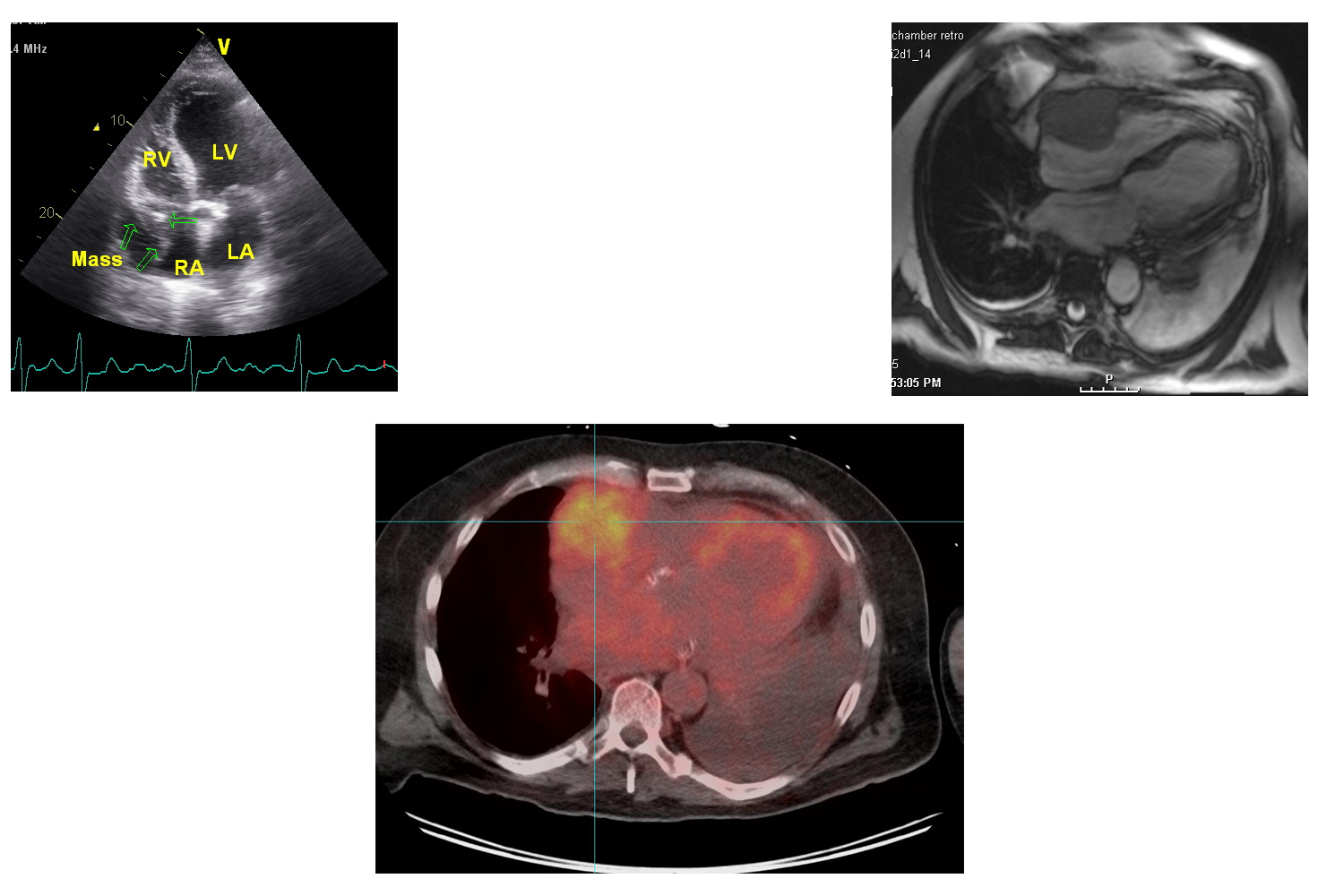
Multiple Myeloma (MM) is a plasma cell dyscrasia characterized by the presence of ‘M’ component in serum and urine along with anemia, hypercalcemia, bony lesions and renal failure. A 76-year-old male with multiple myeloma was admitted with palpitation and dyspnea. The patients had completed chemotherapy and stem cell transplantation for MM with complete remission. On examination the patient had tachycardia with irregular pulse. Pertinent laboratory findings were hemoglobin 8.8 g%, serum potassium 5.6 mg/dl, creatinine 3.8 mg/dl, uric acid 14.2 mg/dl suggestive of tumor lysis. Protein electrophoresis was positive for M band (2.3 g/dl). ECG revealed atrial fibrillation. A transthoracic echo revealed a large, echogenic, spherical, heterogenous fixed mass 63 x 55 mm in the right atrium (Figure 1). Cardiac MRI revealed a mass involving the free wall of the right atrium/ventricle and extending to involve the atrio-ventricular groove (Figure 2). On PET/CT, the mass was FDG avid with SUV 6.0, along with lesions suggestive of new extramedullary involvement of MM (Figure 3). A cardiac biopsy could not be done as the patient declined further therapy. Although atrial myxoma can be a differential diagnosis, it is uncommon in right atrium and rarely shows significant FDG activity [1]. Angiosarcoma, another differential diagnosis, is usually seen as a cardiac mass along with extensive pericardial involvement and hemorrhagic pericardial effusion [2]. Based on intense FDG avidity (SUV 6.0) and the co-existence of other extramedullary lesions, the right atrial mass is likely due MM in the given case.
References
Corresponding author
Jawahar L. Mehta
Division of Internal Medicine
University of Arkansas for Medical Sciences
Little Rock
USA
 Awards Nomination