PHONE
+44-7482-878921
+44-7482-878921
2376-0249
Clinical Image - International Journal of Clinical & Medical Images (2015) Volume 2, Issue 1

Author(s): Alessandro Ginori*c
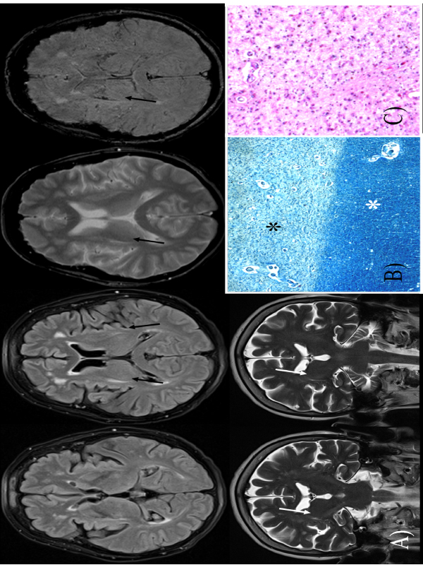
A 60-year-old woman affected by Parkinson disease died for respiratory failure. Before the death, a brain Magnetic Resonance (MR) with contrast media was performed, showing a stria of altered signal in the posterior portion of right putamen (a so called “slit lesion”) (arrows; Figure A). At autopsy, the brain appeared grossly edematous and atrophic. On histologic examination, the Putamen showed severe loss of myelin and axons (Klüver-Barrera myelin-specific stain; black asterisk), while the adjacent cerebral parenchyma appeared well preserved (white asterisk) (40x; Figure B). At higher magnification, the area was characterized by severe demyelination and increase of microglial cells with accumulation of phagocytized material (haematoxylin and eosin, 200x; Figure C).
The presence of a well-demarcated area of atrophy and demyelination suggested the diagnosis of multiple system atrophy (MSA) rather than Parkinson disease. MSA is a neurodegenerative disorder characterized by autonomic failure associated with parkinsonism, cerebellar ataxia, or both, and by impaired sympathetic vasomotor control, manifested with orthostatic hypotension, cardiovagal impairment and respiratory abnormalities. These clinical manifestations are more frequent and severe than in Parkinson disease and were all of them present in our patient. This case is interesting for the high concordance between MR and histology and for the potential clinical meaning of lesions such as that described herein in patients with Parkinson disease.
 Awards Nomination