PHONE
+44-7482-878921
+44-7482-878921
2376-0249
Case Blog - International Journal of Clinical & Medical Images (2016) Volume 3, Issue 7

Author(s): Mahesh Shivaji Chavan, Anagha Shete and Digamber Sable
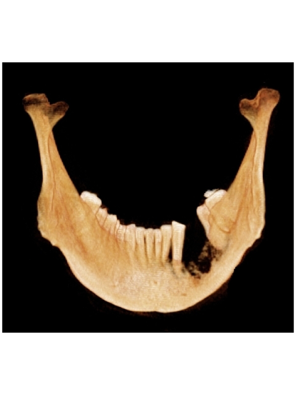
Case Presentation: A 58-year-old male patient was referred to department of oral medicine and radiology for investigations of oral squamous cell carcinoma in left mandibular region. CBCT (cone beam computed tomography) scan of patient revealed bilateral bifid condyle (Figure 1) which was an incidental finding. Patient had no complaints for loss of function, pain or tenderness and clicking sounds associated with both temporomandibular joints. The bifid mandibular condyle (BMC) is a rare anomaly whose cause is not yet fully understood [1]. It can be symptomatic or diagnosed incidentally on routine radiographic examination. No definite etiologic factor has been identified. It is suggested that bifid condyle could be a developmental anomaly or secondary to trauma [2]. CT (computed tomography) scan is the best radiograph for detection of BMC because it allows for detailed evaluation of condylar morphology. However, BMC can also be seen on OPG, but sometimes the overlapping of the anatomic structures can hide the bifidity [2]. Tanner et al. [3] suggested that, 3D CT reconstructed images are useful to accurately diagnose BMC as well as to fully assess whether the condylar heads emerge from the mandibular neck. CBCT has less radiation exposure to patients than CT scan so it is advised to use CBCT scans in oral and maxillofacial region.
 Awards Nomination