PHONE
+44-7482-878921
+44-7482-878921
2376-0249
Clinical Image - International Journal of Clinical & Medical Images (2014) Volume 1, Issue 2

Author(s): Hussein Abujrad MBBCh, FRCPC, Heather Lochnan MD
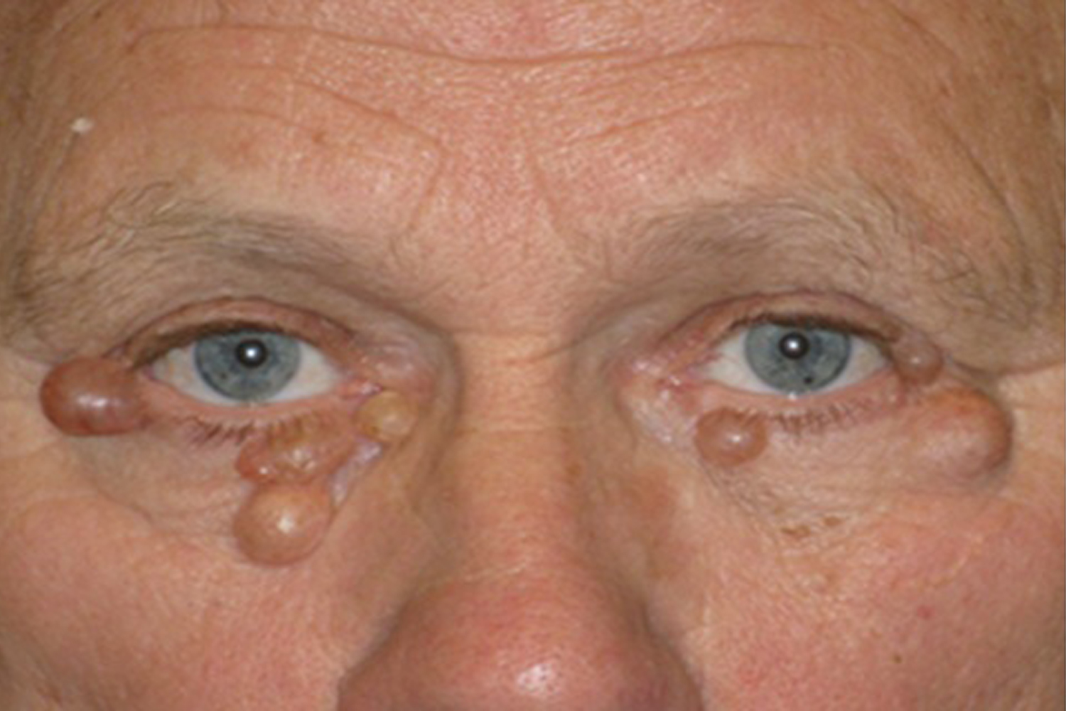
A 67 year-old male presented with bilateral painless periorbital lesions that appeared to be cystic. These lestions became apparent one year prior and were progressing in size. He described having similar lesions removed many years previously, but they were not as dramatic at that time (Figure 1). His medical history was significant for hypogonadism and hyperprolactinemia recently diagnosed and found to be secondary to a pituitary microadenoma (Microprolactinoma) and he is known to have significant coronary artery disease. His family history was unremarkable. The patient was referred to the dermatological assessment and Hidrocystoma (Eccrine) was diagnosed and treated with surgical drainage. Hidrocystomas are benign, cystic, sweat gland tumours of the skin that occur mostly in adults between 30 and 70 years of age. They are classified as either apocrine or eccrine. Apocrine hidrocystomas arise from the proliferation of apocrine glands and are usually solitary, with a diameter of 3–15 mm. Eccrine hidrocystomas result from cystic dilation due to retention of sweat and blockage of the sweat duct. They are tense, dome-shaped cysts, ranging from 1 to 6 mm in diameter and usually affect the periorbital and malar areas but do not involve the eyelid margin. They may have an amber, brown, or bluish tint. Our patient has no recurrence of the hidrocystomas after 10 months of the surgical drainage and no obvious connection to his prolactinoma can be made.
Corresponding authors
Hussein Abujrad and Heather Lochnan
Division of Endocrinology and Metabolism
University of Ottawa
Canada
 Awards Nomination