PHONE
+44-7482-878921
+44-7482-878921
2376-0249
Clinical-Medical Image - International Journal of Clinical & Medical Images (2024) Volume 11, Issue 2

Author(s): Muhammad Arif Ozir*
Department of Ophthalmology, Hospital Pengajar Universiti Sultan Zainal Abidin, Kuala Terengganu, Malaysia
*Corresponding Author:
Muhammad Arif Ozir
Department of Ophthalmology
Hospital Pengajar Universiti Sultan Zainal Abidin
Kuala Terengganu, Malaysia
E-mail: drmuhammadarif@yahoo.com
Received: 01 February 2024, Manuscript No. ijcmi-24-119800; Editor assigned: 03 February 2024, Pre QC No. P-119800; Reviewed: 15 February 2024, QC No. Q-119800; Revised: 21 February 2024, Manuscript No. R- 119800; Published: 29 February 2024, DOI:10.4172/2376-0249.1000944
Citation: Ozir MA. (2024) Branch Retinal Vein Occlusion. Int J Clin Med Imaging 11: 944.
Copyright: © 2024 Ozir MA. This is an open-access article distributed under the terms of the Creative Commons Attribution License, which permits unrestricted use, distribution, and reproduction in any medium, provided the original author and source are credited.
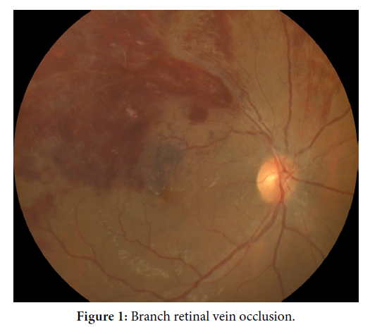
A 38-year-old man with comorbidity of diabetis mellitus,hypertension and hyperlipidemia presented to the ophthalmology clinic with right eye painless progressive loss of vision for 1 month. During presentation, vision was 6/36 OD and 6/6 OS. Anterior segment ophthalmic examination of bilateral eyes is normal. Fundus examination of right eye showing superotemporal branch retinal vein occlusion. There is classic flame shape hemorrhage and cotton wool spots. The vein is tortuous and dilated. In area of flame shape haemorrhage there is sclerosed vein in between. Ocular Coherence Tomography (OCT) macula show macula edema. A diagnosis of branch retinal vein occlusion was made. BRVO is a venous occlusion at any branch of the central retinal vein. The incidence of BRVO is most common in the superotemporal quadrant (58.1 - 66%), followed by the inferotemporal quadrant (29%), and least common in the nasal quadrants (12.9%). The higher incidence of superotemporal BRVO compared to other quadrants is speculated to be due to a larger number arteriovenous crossings in that quadrant, which supports the hypothesis of arteriovenous nicking as a cause. This patient is scheduled for anti-VEGF injection due to macula edema. He was also advised for strict control of blood pressure, glucose and cholesterol due to his comorbidity [1,2].
Ocular Coherence Tomography (OCT); Diabetis mellitus
None.
[1] Kim CS, Shin KS, Lee HJ, Jo YJ and Kim JY. (2014). Sectoral retinal nerve fiber layer thinning in branch retinal vein occlusion. Retina 34(3): 525-530.
Google Scholar, Crossref, Indexed at
[2] Vernazza S, Oddone F, Tirendi S and Bassi AM. (2021). Risk factors for retinal ganglion cell distress in glaucoma and neuroprotective potential intervention. Int J Mol Sci 22(15): 7994.
Google Scholar, Crossref, Indexed at
 Awards Nomination