PHONE
+44-7482-878921
+44-7482-878921
2376-0249
Clinical-Medical Image - International Journal of Clinical & Medical Images (2022) Volume 9, Issue 5

Author(s): Abdelilah Drissi*, A Maniani, Habibchorfa Sara, Abir Lemrabet Meriem Fikri, Mohamed Jiddane and Najwa Ech-Chrif El Kettani
Received: 04 May 2022, Manuscript No. ijcmi-22-65951; Editor assigned: 06 May 2022, Pre QC No. P-65951; Reviewed: 17 May 2022, QC No. Q-65951; Revised: 19 May 2022, Manuscript No. R-65951; Published: 27 May 2022, DOI: 10.4172/2376-0249.1000828
Clinical-Medical Image
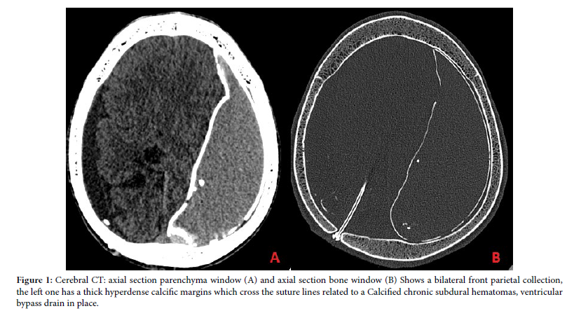
Our case is about 18 year old male, with a history of hydrocephalus with a ventriculo-peritoneal shunt during his childhood, who was admitted to the emergency room for seizures. A cerebral CT was performed who shows a bilateral collection of the cerebral convexities, the left one has a thick hyperdense calcific margins characteristic of a chronic calcified subdural hematoma (Figure 1).
Discussion
The Chronic calcified subdural hematoma (HSDC) is arare entity, it presents 0.3 to 2.7% of chronic subdural hematomas, more frequently in children and young adults. It is a collection of blood between the arachnoid and the dura mater, the interval between the acute phase and the appearance of calcification varies from 6 months to several years [1,2]
Clinically it shares the same signs with chronic HSDs (seizures, focal neurological deficits or mental retardation) but may be asymptomatic. Standard X-ray of the skull shows a calcified lesion that fits the curvature of the skull. CT brain shows a hypodense extra-axial collection typically appears as a crescent-shaped with thick and calcified margins which cross the suture lines and exert a variable mass effect. A bilateral location gives a “double skull” appearance called the “Matrioska head”. MRI is less sensitive than CT in detection of a calcified mass but it has an interest for evaluating the degree of adhesion of calcification to the underlying structures. The neurosurgical intervention is discussed case by case, depending on the prognosis and the degree of adhesion of the calcification to the underlying structures [3].
Final diagnosis
Calcified chronic subdural hematoma
Three differential diagnosis:
• Calcified epidural hematoma
• Meningioma Calcified empyema
Keywords: Malignant tumor; Chronic calcified subdural hematoma; MRI; HSDC
References
[1] Pappamikail L, Rato R, Novais G, Bernardo E (2013). Chronic calcified subdural hematoma: Case report and review of the literature. Surg Neurol Int 4.
GoogleScholar Crossref Indexed at
[2] Niwa J, Nakamura T, Fujishige M, Hashi K (1988) Removal of a large asymptomatic calcified chronic subdural hematoma. Surg Neurol 30(2) : 135-139.
GoogleScholar Crossref Indexed at
[3] Ide M, Jimbo M, Yamamoto M, Umebara Y, Hagiwara S (1993). Asymptomatic calcified chronic subdural hematoma-Report of three cases. Neurol. Med Chir 33(8): 559-563.
 Awards Nomination