PHONE
+44-7482-878921
+44-7482-878921
2376-0249
Clinical-Medical Image - International Journal of Clinical & Medical Images (2022) Volume 9, Issue 2

Author(s): Sharmistha Chakravarty*, Nitin M Nagarkar, Rupa Mehta, Ripu Daman Arora
Received: 01 February, 2022, Manuscript No. ijcmi-22-53937; Editor assigned: 02 February, 2022, PreQC No. P-53937; Reviewed: 17 February, 2022, QC No. Q-53937; Revised: 22 February, 2022, Manuscript No. R-53937; Published: 28 February, 2022, DOI: 10.4172/2376-0249.1000814
Clinical-Medical Image
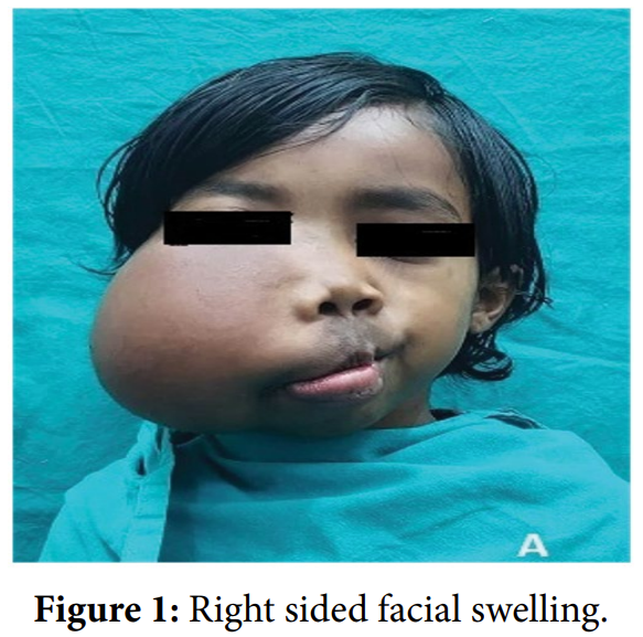
A 6 year old girl presented to the Otolaryngology clinic with complaint of right sided facial swelling since birth which has progressively increased in size leading to facial asymmetry. It was a diffuse non-tender swelling, soft in consistency and extended from zygotic arch above to right hemimandible below, medially extending up to midline of nose and mouth and laterally up to the parotid region of the right side. There was history of early loss of deciduous teeth and eruption of permanent teeth on the right side of upper and lower jaw. There was no complaint of pain, hypoaesthesia, localised muscle weakness or throbbing sensation. On examination, the swelling was approximately 10 × 8 cm right sided facial swelling, soft on palpation, overlying skin was warm to touch and the colour and texture of the skin was within normal limits (Figure 1). Oral cavity examination showed enlarged teeth with hypertrophy of right alveolar arch and buccal mucosa (Figure 2). USG showed an illdefined; heterogeneously hypoechoic lesion measuring 10 × 15 cm and no vascular anomaly was noted. MRI of face showed TI hyperintense cheek mass with multiple septations, atrophy of muscles of right side of face with fatty infiltration of masseter (Figure 3) and temporalis muscle, associated with fatty hypertrophy of soft tissue plane showing fat suppression on STIR images (Figure 4). There is mucosal thickening of paranasal sinuses with widening of intramedullary region of anterior wall of right maxilla. CT scan of face showed mild enlargement of right maxilla and mandible, zygomatic and sphenoid bone. The radiological findings were compatible with Congenital Infiltrative Lipomatosis of face. Vascular malformations or haemangioma was ruled out based on USG, CT and MRI images. FNAC from the mass revealed clusters of mature adipocytes on a greasy background with no atypical cells favouring the diagnosis to be a benign lipomatous lesion. Congenital Infiltrating Lipomatosis of the face (CILF) is a very rare disorder in which mature lymphocytes invade into adjacent soft and bony tissues in the facial region. The gradual enlargement of the affected side of the face can lead to facial asymmetry, macrodontism, early eruption of deciduous and permanent teeth, and hypertrophy of bones, macroglossia, and the proliferation of parotid gland of the affected side. The patient underwent a surgical excision of the mass by subcutaneous lipectomy which was done by multidisciplinary team comprising of otolaryngologist, plastic surgeon and maxillofacial surgeon. The patient has recovered well with intact facial nerve function and is on a six month follow up without recurrence of growth [1].
Keywords: Congenital; Lipomatosis; Facial asymmetry; Macrodontism
Declaration of Conflicting Interests
The authors declared no potential conflicts of interest with respect to the research, authorship, and/or publication of this article.
References
[1] Touimi SH, Mbarki I, Elkacemi H, Elmajjaoui S, Kebdani T, et al. (2020) An Atypical Case of Epithelial-myoepithelial Carcinoma of the Lacrymal Gland. Int J Clin Med Imaging 7: 676
 Awards Nomination