PHONE
+44-7482-878921
+44-7482-878921
2376-0249
Clinical Image - International Journal of Clinical & Medical Images (2014) Volume 1, Issue 6

Author(s): Dan Xia, Shuai Zhang, Jesper ?stergaardHjortdal and Mingdong Dong
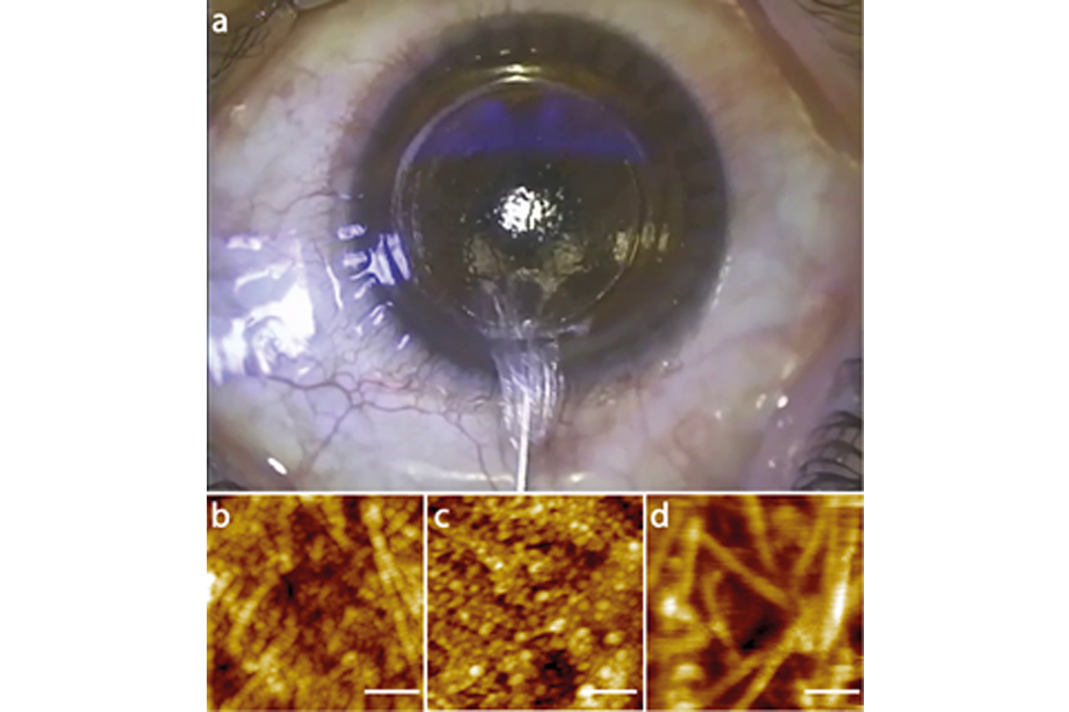
Small incision lenticule extraction (SMILE) is a novel form of ‘flapless’ corneal refractive surgeryfor remodeling the profile of the cornea by removing the stroma to correct myopia (Figure a). The stromas prepared by the SMILE surgery under different hydration levels are studied by atomic force microscopy (Figure b-d). It shows the morphologies are significantly related to the hydration levels. The stroma in 40% humidity (Figure b) shows fine collagen fiber with clear periodicity. However, the stroma immersed in physiological buffer for 0.2h (Figure c) show particle protrusions along the fiber and the periodicity disappears. The longer time immersed stroma in physiological buffer (1h) shows fiber with bigger width, and again no periodicity can be seen (Figure d). This phenomenon indicates that the stroma swells when it is immersed in physiological buffer. This proves the stroma has an innate tendency to imbibe fluid and its biophysical property depends on precisely maintaining its hydration level. (a) The photo captured during the refractive lenticule extraction SMILE surgery; (b) Morphology of the corneal stroma in 40% humidity; (c) Morphology of the corneal stroma immersed in physiological buffer for 0.2 hour; (d) Morphology of the corneal stroma immersed in physiological buffer for 1 hour. All the scale bars are 250 nm.
*Corresponding author: Mingdong Dong, Interdisciplinary Nanoscience Center (iNANO), Aarhus University, 8000 Aarhus C, Denmark, Tel: 87156729, E-Mail: dong@inano.au.dk
 Awards Nomination