PHONE
+44-7482-878921
+44-7482-878921
2376-0249
Case Blog - International Journal of Clinical & Medical Images (2015) Volume 2, Issue 3

Author(s): Artul S*, Nseir W, Yamini A, Shkara HA, Joubran H and Habib G
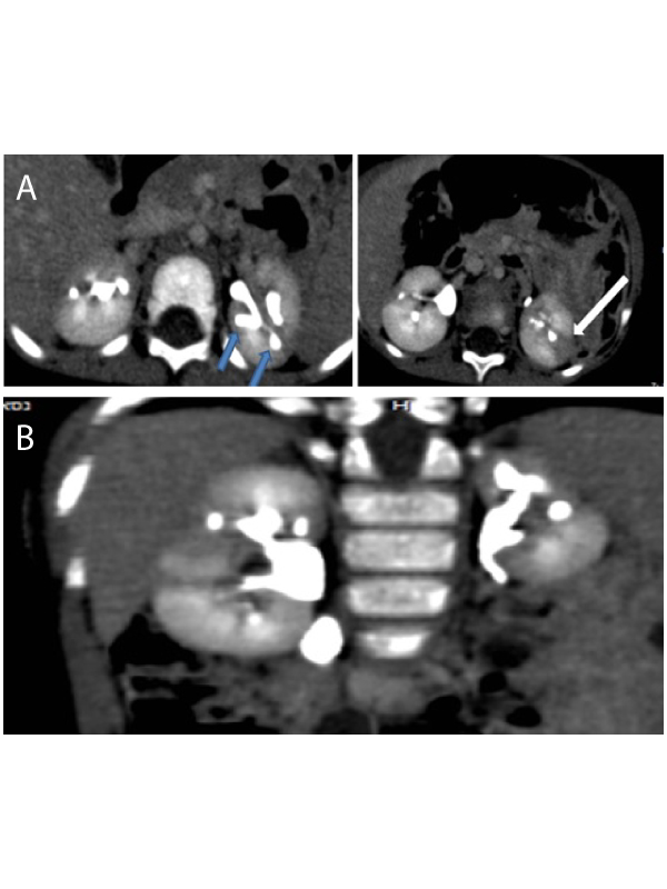
Clinical Image: Two year old infant admitted to the emergency department after severe back injury. Urine test was full of blood. CT of abdomen showed multiple hypo-dense lesions in both kidneys resembling hematomas (Figure 1), dilated collecting systems, lobulation of the outer contour of both kidneys and small left kidney (Figure 2). There were no extravasations and no fat infiltration around the kidneys. These signs were compatible with intrarenal reflux to the kidneys and not traumatic findings. During review with the family of the past medical history it was discovered that this infant is in follow up for recurrent urinary tract infection and vesicoureteral reflux (VUR) in another hospital. VUR may be asymptomatic and resolve spontaneously during childhood without changes to the upper urinary tracts or may persist and cause permanent damage to the kidneys. Children with VUR are predisposed for recurrent urinary tract infections [1].
VUR are classified into 5 grades Persistent VUR grade 4 and 5 (VUR with moderate/severe dilatation to collecting system), this may cause renal parenchymal scarring [2]. Intrarenal reflux is the most severe grade of reflux (reflux to the parenchymal collecting tubules) and when this is present; thekidney will usually develop scarring and atrophy. Scarring may involve a very small portion of a kidney or nearly the entire organ. The scarring has a predilection to appear in the poles, especially the upper poles [1,2]. CT signs of intrarenal VUR are: Scarring that appears as hypo-dense lesions without enhancement, dilated collecting system, Clubbing of the calyces (Figure 1) and lobulated outer contour of the kidney
 Awards Nomination