PHONE
+44-7482-878921
+44-7482-878921
2376-0249
Clinical-Medical Image - International Journal of Clinical & Medical Images (2023) Volume 10, Issue 5

Author(s): Siham Oukassem*, Abourak Chaimae, Jihan El Houssni, Ilyass Bourekba, Meryem Eddrai, Hassan Ennouillia and Jamal El Fenni
Department of Radiology, Mohammed Vth Military Hospital, Ryad Street, 10010 Rabat, Morocco
Received: 08 April 2023, Manuscript No. ijcmi-23-94858; Editor assigned: 10 April 2023, Pre QC No. P-94858; Reviewed: 24 April 2023, QC No. Q-94858; Revised: 01 May 2023, Manuscript No. R-94858; Published: 08 May 2023, DOI:10.4172/2376-0249.1000892
Citation: Oukassem S, Chaimae A, Houssni JEI, Bourekba I and Eddrai M, et al. (2023) Cutaneous Metastasis Revealing Adrenocortical Carcinoma: A Case Report. Int J Clin Med Imaging 10:892.
Copyright: © 2023 Oukassem S, et al. This is an open-access article distributed under the terms of the Creative Commons Attribution License, which permits unrestricted use, distribution, and reproduction in any medium, provided the original author and source are credited.
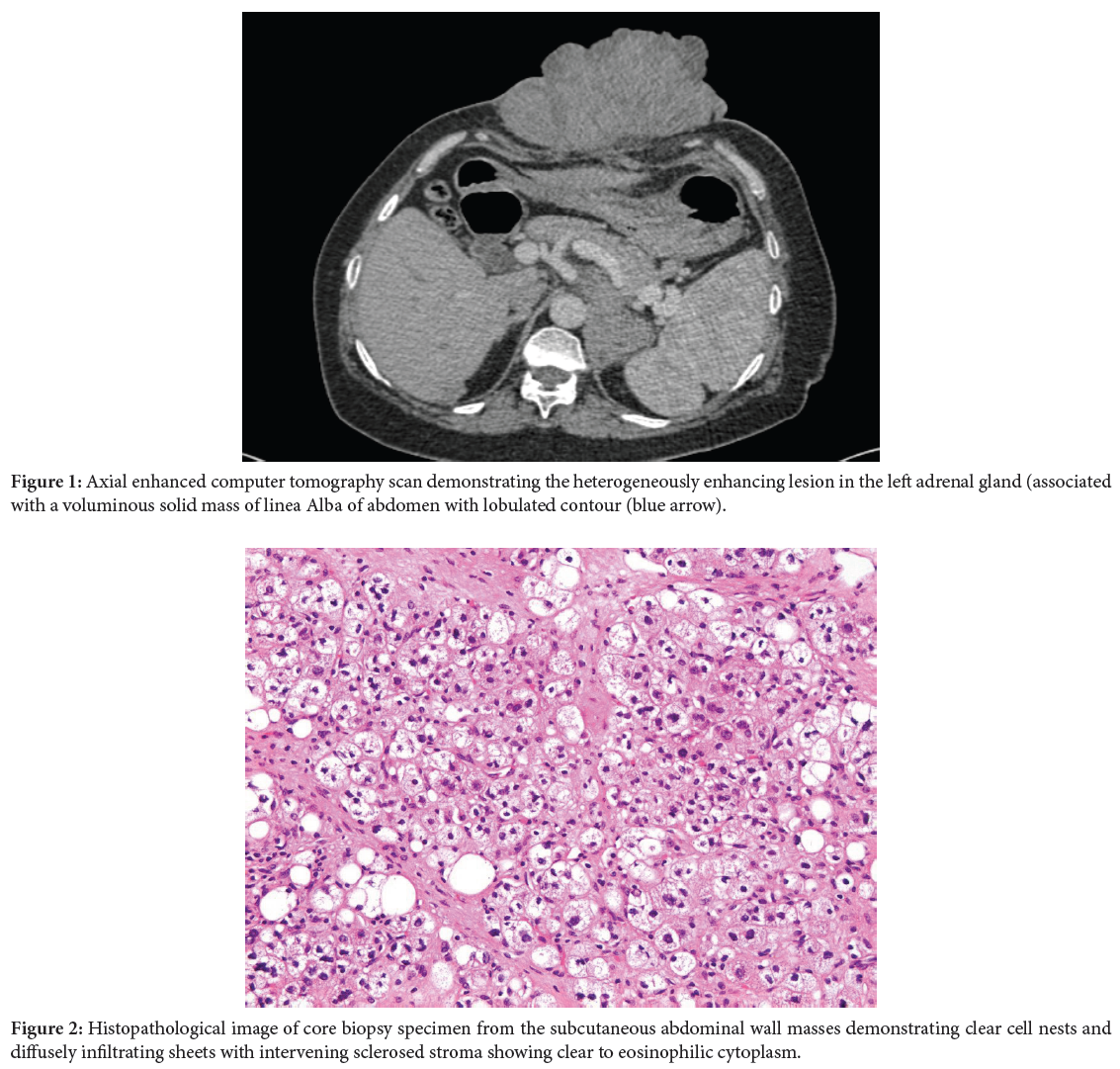
We report the case of 50 year a -old female consulted for a cutaneous lesion in abdominal wall, whose exploration had shown the existence of a large adrenal lesion. Axial enhanced computer tomography scan images (Figure 1) demonstrating the heterogeneously enhancing lesion in the left adrenal gland with areas of necrosis associated with a voluminous mass solid mass of linea alba of abdomen. Histologic comparison proved that the cutaneous lesion was a metastase from an adrenocortical carcinoma (Figure 2).
Adrenocortical carcinomas are very aggressive lesions and in some cases may be functional and present with Cushing syndrome and/or virilization. In most cases, ACC is non-functional and presents as an abdominal mass or an incidental finding [1]. Size >4 cm, rapid growth, heterogeneous shape, irregular borders, central necrosis, hemorrhage, calcification, invasion into neighboring structures and venous extension are imaging findings indicative of adrenal cancer. 70% of all ACC are generally >6 cm and can reach a maximum size of 25 cm. Systemic or local metastasis is possible. The pancreas, spleen, liver, gut, retroperitoneum, venous extension to the inferior vena cava and lymphatic dissemination through regional and para-aortic lymph nodes are typical sites of local metastasis. Systemic metastases occur most frequently in the lung (40%-80%), liver (40%-90%), bone (5%-20%), Inferior vena cava (9%-19%) and brain and skin (<5%) [2]. Despite a high frequency of metastases, there are only isolated reports of ACC that have metastasized to the skin. On unenhanced CT, the ACC appears large, poorly defined, heterogenous, with HU >10 and areas of calcification and necrosis. In contrast-enhanced CT scans, the tumor appears heterogeneously enhanced with significant peripheral enhancement because of the core necrosis (3). CECT is the only reliable method for staging and identifying metastatic sites [3]. The nine parameters of the histopathological analysis used to determine prognosis and distinguish benign from malignant adrenal cortical tumors by the Weiss score include nuclear grading, mitotic rate of >5/50 HPFs, abnormal mitoses, 25% clear cells, >1/3 diffuse architecture, necrosis, venous invasion, sinusoidal invasion and capsular invasion [4].
Adrenocortical carcinoma; Cutaneous metastasis; CT scan
None of the authors has any conflicts of interests to disclose.
[1] Rodgers SE, Evans DB, Lee JE and Perrier ND. (2006). Adrenocortical carcinoma. Surg Oncol Clin N Am 15: 535-53.
Google Scholar, Crossref, Indexed at
[2] Fishman EK, Deutch BM, Hartman DS, Goldman SM and Zerhouni EA, et al. (1987). Primary adrenocortical carcinoma: CT evaluation with clinical correlation. AJR Am J Roentgenol 148: 531-535.
Google Scholar, Crossref, Indexed at
[3] Dunnick NR, Heaston D, Halvorsen R, Moore AV and Korobkin M, et al. (1982). CT appearance of adrenal cortical carcinoma. J Comput Assist Tomogr 6: 978-982.
Google Scholar, Crossref, Indexed at
[4] Jain M, Kapoor S, Mishra A, Gupta S and Agarwal A. (2010). Weiss criteria in large adrenocortical tumors: A validation study. Indian J Pathol Microbiol 53: 222.
 Awards Nomination