PHONE
+44-7482-878921
+44-7482-878921
2376-0249
Clinical-Medical Image - International Journal of Clinical & Medical Images (2023) Volume 10, Issue 3

Author(s): Younes Akannour*, El Akhdari Meryem, Louai Serghini, Abdallah Elhassan and Berraho Amina
Department of Ophthalmology B, Hospital of Specialties of Rabat (CHU Ibn Sina), Mohammed V University, Morocco
Received: 15 March 2023, Manuscript No. ijcmi-23-91865; Editor assigned: 16 March 2023, Pre QC No. P-91865; Reviewed: 30 March 2023, QC No. Q-91865; Revised: 05 April 2023, Manuscript No. R-91865; Published: 12 April 2023, DOI:10.4172/2376-0249.1000885
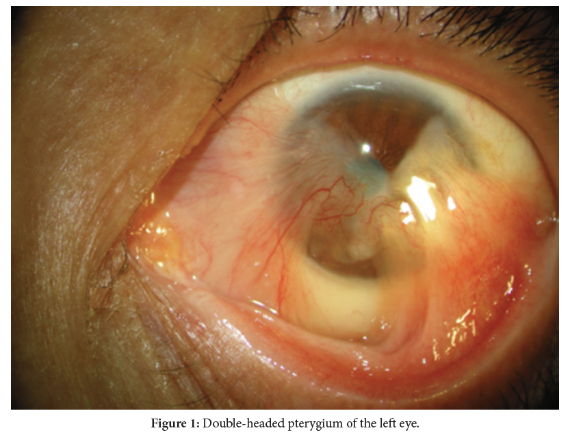
Citation: Akannour Y, Meryem EA, Serghini L, Elhassan A and Amina B. (2023) Double-Head Pterygium Treated with Amniotic Membrane Graft. Int J Clin Med Imaging 10:885.
Copyright: © 2023 Akannour Y, et al. This is an open-access article distributed under the terms of the Creative Commons Attribution License, which permits unrestricted use, distribution and reproduction in any medium, provided the original author and source are credited.
A pterygium is a benign conjunctival fibrovascular neoformation, characterized by its recurrent and progressive nature in the direction of the cornea. Double-headed pterygia are rare and present the surgeon with challenges. We report the case of 53-year-old man was referred to our eye hospital with a double-head pterygium of the right eye. The patient had hand movement visual acuity on the right eye, slit-lamp examination foud that the head of the pertygium encroaching towards the center of the cornea on both sides. After local anesthesia, the pterygium with subconjunctival fibrovascular tissue is removed. The area of corneal scar is carefully smoothed and denuded. Bipolar cautery is used to maintain hemostasis. One piece of amniotic membrane is placed on cornea, the bare sclera and under the large conjunctival flap, the grafts were sutured using interrupted 10-0 nylon sutures, we employ the amniotic membrane graft to have antifibrotic and anti-inflammatory effects, this application facilitates also epithelialization at the area of the corneal defect after pterygium excision. We chose to use an amniotic membrane rather than autografts because the remaining part of the conjunctiva was too small and insufficient. Post operatively the patient received a single intravenous dose of methylprednisolone. He was prescribed topical steroids 6 times a day for a month and artificial tears [1,2].
Double-head pterygium; Pterygium; Amniotic membrane
The authors received no financial support for the research, authorship, and/or publication of this article.
None of the authors has any conflicts of interests to disclose.
[1] Bhatia S, Gupta A, Sachdev R and Bhatia R. (2014) Management of double-head pterygium with autologous limbal conjunctival grafting. Oman J Ophthalmol 7: 144–146.
Google Scholar, Crossref, Indexed at
[2] Kocak-Altintas AG, Karaman E, Duman R and Kocabeyoglu S. (2016). Double-head pterygium: Case report and review of the literature. Turkish J Ophthalmol 46: 90–93
 Awards Nomination