PHONE
+44-7482-878921
+44-7482-878921
2376-0249
Medical Image - International Journal of Clinical & Medical Images (2016) Volume 3, Issue 2

Author(s): Shalabh Kumar A, Sohan Kumar S, Kapil B and Mukesh
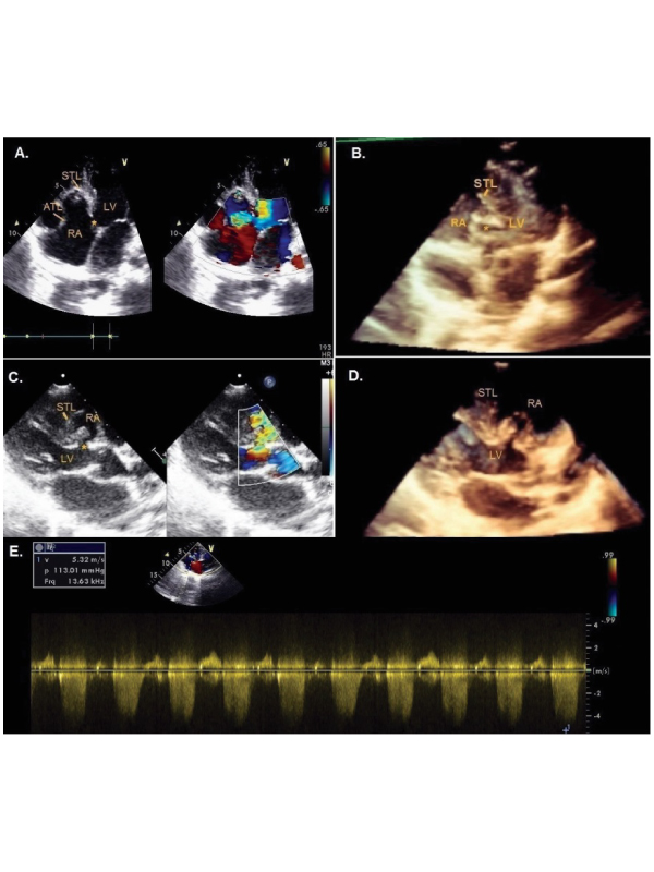
Panel A: Two-dimensional and colour flow TTE from Apical four chamber view showing apically displaced STL and sail like ATL with a defect in ventriculoatrial septum (*) with LV to RA flow being simultaneously demonstrated.
Panel B: Three-dimensional TTE showing the same defect.
Panel C: Two-dimensional TTE from Parasternal long axis view demonstrating the defect (*) with colour flow image confirming the presence of highly turbulent jet from LV to RA. Panel D: Three-dimensional TTE which clearly depicts the defect.
Panel E: Continuous Wave Doppler demonstrating a high peak instantaneous gradient of 113 mm Hg across the defect.
A 7 year old boy underwent transthoracic echocardiography (TTE) for evaluation of effort intolerance and frequent palpitations with a harsh pansystolic murmur audible all over the precordium. TTE revealed the presence of Ebstein’s anomaly with apically displaced septal tricuspid leaflet (STL) and ‘sail like’ anterior tricuspid leaflet (ATL). There was a direct ventriculoatrial shunt from Left ventricle (LV) to Right atrium (RA) –Gerbode defect with a peak systolic gradient of 113 mm Hg. Ebstein’s anomaly is commonly associated with various other congenital heart defects but association of Ebstein’s with Gerbode defect is very rare (Panel A-E).
 Awards Nomination