PHONE
+44-7482-878921
+44-7482-878921
2376-0249
Clinical-Medical Image - International Journal of Clinical & Medical Images (2021) Volume 8, Issue 2

Author(s): Kaoutar Imrani, Belkouchi Lina, Oubaddi Tlaite, Hounayda Jerguigue, Rachida Latib, and Youssef Omor
Clinical Image
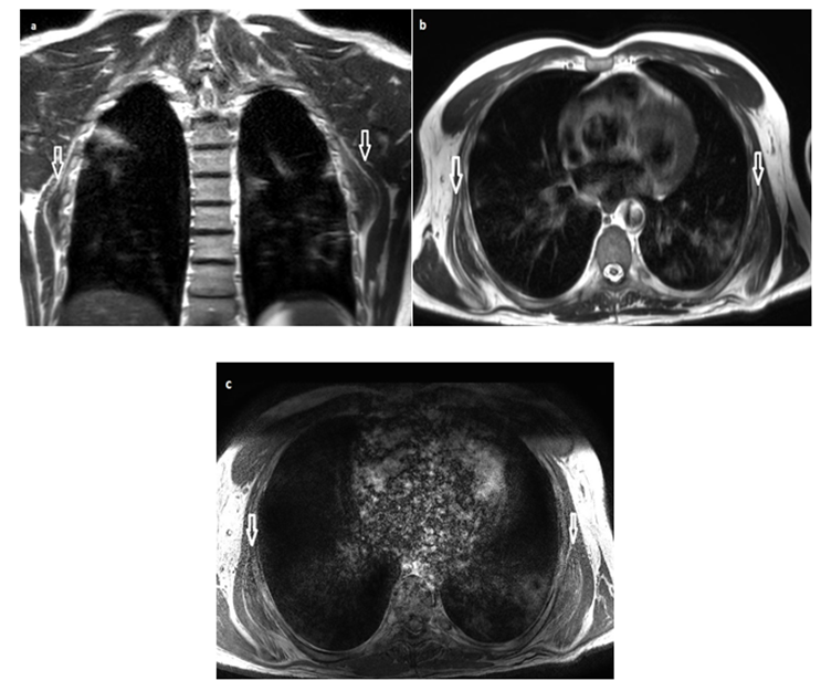
Elasto-fibroma dorsi is a benign tumour first described by Jarvi and Saxen in 1959. It is most often located in the sub-and pre-scapular region [1]. Clinically, elastofibroma is often asymptomatic, but may generate moderate scapular pain or a limitation of arm abduction. Imaging has an important role in diagnosis. The ultrasound shows a very limited lesion, with alternating hypoechogenic linear layers corresponding to the fatty tissue, and echogenic linear layers corresponding to the elastic fibro tissue. CT scans shows a sub- or sub-scapular mass, hypodense, striated, similar to adjacent muscle structures [2]. MRI provides better characterization of the lesion presenting as a well-defined mass, not encapsulated, alternating layers of fibrous tissue iso intense to muscle in T1 and T2-weighted sequences, and layers of fat tissue hyperintense in T1 and T2, not suppressed after fat saturation, enhanced moderately and heterogeneously after gadolinium injection, taking a thousand leaves appearance (Figure 1). The differential diagnosis arises mainly with desmoid tumors, neurofibromas and liposarcoma. The treatment is based on surgical excision, it is indicated especially when the tumour is voluminous, painful or in case of aesthetic discomfort [1,2].
Keywords: Elastofibroma; MRI; Thousand leaves
 Awards Nomination