PHONE
+44-7482-878921
+44-7482-878921
2376-0249
Case Blog - International Journal of Clinical & Medical Images (2014) Volume 1, Issue 11

Author(s): IAlexander K. C. Leung* and Benjamin Barankin
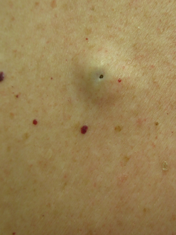
A 56-year-old woman presented with an asymptomatic, slowly growing, dome-shaped mass on the back of four years’ duration. There was no known history of local trauma or infection. On examination, a firm, nontender, mass was found on the back. The mass was not attached to any underlying structure. An epidermoid cyst is filled with keratin flakes or debris and its wall is composed of keratinized stratified squamous epithelium. Most cases are acquired and secondary to traumatic or iatrogenic implantation of epithelial cells into the dermal or subcutaneous layer or from obstruction of a pilosebaceous unit in the hair follicle. Typically, an epidermoid cyst presents as an asymptomatic, slow-growing, fluctuant to firm, dome-shaped mass that is not attached to the underlying structure. Epidermoid cysts occur mainly on hair-bearing areas. The condition is usually solitary, but uncommonly, can be multiple. Most epidermoid cysts are 1 to 5 cm in diameter and are unilocular. The occurrence is usually sporadic but certain hereditary syndromes such as Gorlin syndrome, Gardner syndrome, and Lowe syndrome have epidermoid cysts as part of their constellation of features. An epidermoid cyst may be cosmetically unsightly and socially embarrassing if it occurs in an exposed area. The cyst may rupture spontaneously or as a result of trauma with release of keratinous material that results in erythema, swelling and pain. There can be an odor emanating from these cysts. An epidermoid cyst may become secondarily infected which can result in abscess formation. Rarely, squamous cell carcinoma, basal cell carcinoma, Bowen’s disease, melanoma, and mycosis fungoides may develop in an epidermoid cyst. If removal of an epidermoid cyst is desired for cosmetic purposes or complications, complete excision of the cyst contents and cyst wall is the treatment of choice. Incomplete excision may lead to chronic inflammation and recurrence. Intralesional cortisone for inflamed and painful cysts is beneficial to bring down the pain and swelling.
 Awards Nomination