PHONE
+44-7482-878921
+44-7482-878921
2376-0249
Clinical-Medical Image - International Journal of Clinical & Medical Images (2019) Volume 6, Issue 6

Author(s): Dagar H* and Norouzbeigi H
Clinical Image:
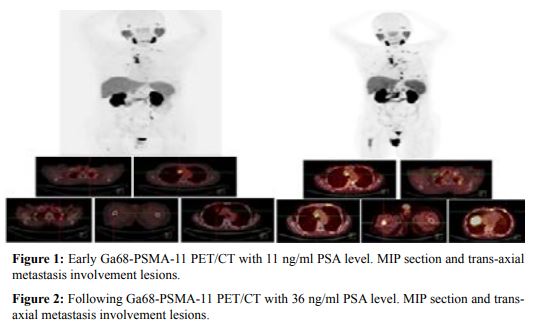
A 74 years old male patient underwent Ga-68 PSMA-HBED-CC PET/CT for restaging of prostate carcinoma. He had diagnosed as prostate adenocarcinoma in 2016 and been followed up in 2017 using Ga-68 PSMA-11. Additionally chemoradiation therapy using taxotere following partial cystectomy had been applied for high grade invasive prostate carcinoma. After chemotherapy and hormone therapy (Zometa), patient underwent Ga-68 PSMA PET/CT for elevation of serum prostate specific antigen levels (early scan). In the early PET/CT scan patient had PSA=11 ng/ml. For this procedure 60 MBq of Ga-68 PSMA-11 was administered intravenously via the left antecubital vein. To allow for distribution and uptake of radiotracer, the patient was allowed to rest quietly for 60 min in a shielded room. Imaging was performed on an integrated 6-slice PET/CT scanner, with scanning from the skull base to the toes. CT scanning was performed without oral or intravenous contrast material. In maximum intensity projection and trans-axial fused images, multiple medistinal lymph node involvement in supraclavicular, restrosternal, lower paratracheal and left internal mammary stations high uptake were detected (Figure 1). In addition, lymph node involvement in the para-aortic area (3-4 small sizes lymph nodes), right iliac wing and T9 and 11th left ribs were reported. After following the patient who underwent hormone therapy each three months, PSA level rose up from 11 to 36 ng/ml and chemotherapy with no change in PSA levels was regarded. Therefore, Ga-68 PSMA PET/CT has been performed again for imaging of recurrent prostate carcinoma progression. After interpretation of these results, we found same scan patterns like pervious study in the bilateral supaclavicular lymph nodes, 9 mm lymph node in the paratracheal region, 15 mm lymph node in the retrosternal area. Furthermore, 2-3 another lymph nodes in the lower paratracheal and retrosternal stations with increased uptake were noted. Lymph node in AP window which was 8 mm with SUV max=10 now became 20 mm with SUV max=17.3. Finally, new lymphatic involvements in the left retrocrural, AP window in addition to the previously noted multiple medistinal lymph node involvement in supraclavicular, retrosternal, lower paratracheal and left internal mammary stations were diagnosed. Moreover, lymph node involvement in the para-aortic area (3-4 small sized lymph nodes) and new bone lesions in the sternum, left scapula, multiple ribs, T5, multiple lower thoracic and lumbar vertebrae and iliac wings were seen (Figure 2).
 Awards Nomination