PHONE
+44-7482-878921
+44-7482-878921
2376-0249
Case Blog - International Journal of Clinical & Medical Images (2014) Volume 1, Issue 7

Author(s): Guillén Morales*, Carolina, Fuerte Ruiz, Sagrario, Carrascosa Mirón and Teresa
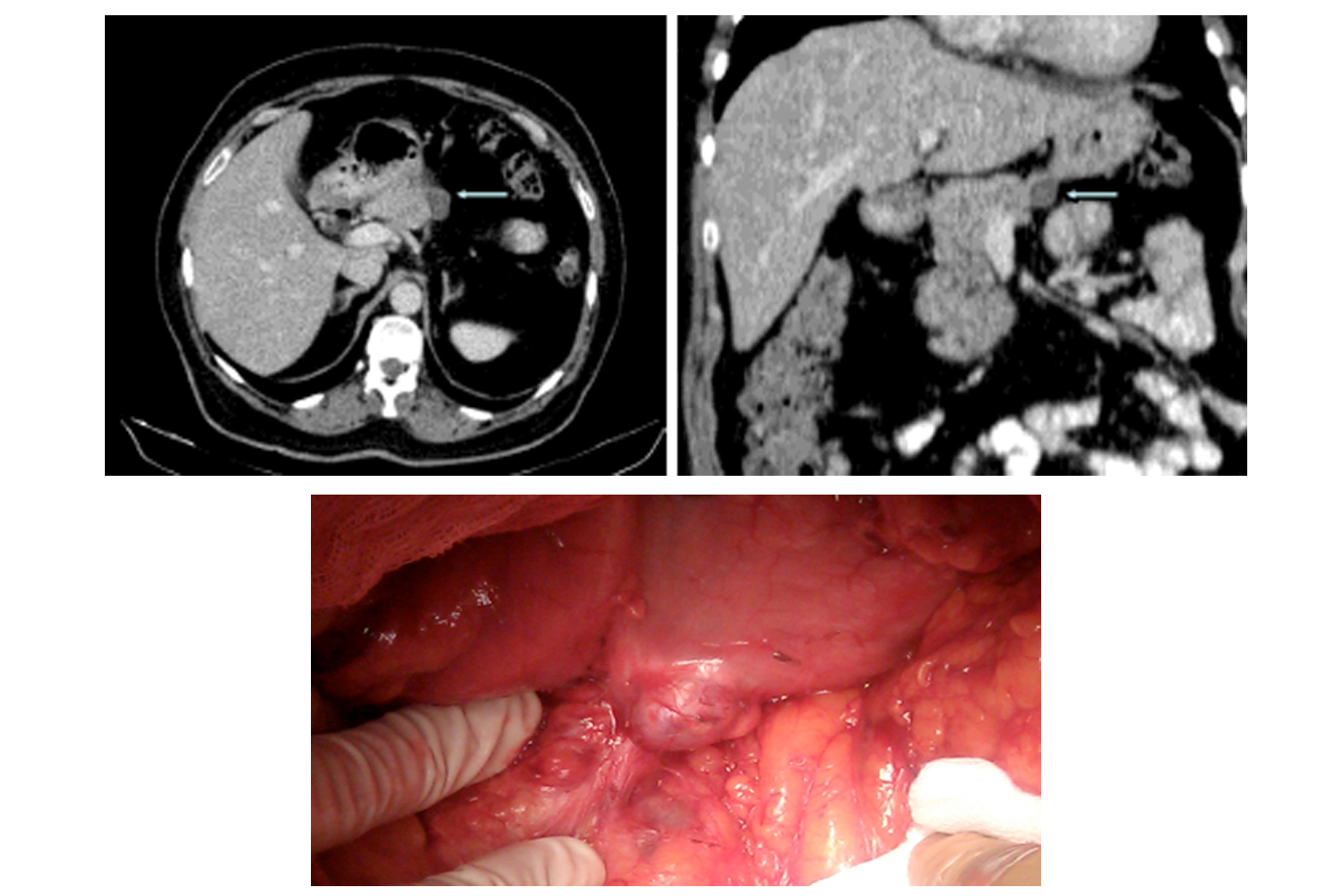
A 55-year-old woman with a history of distal pancreatectomy, cholecystectomy and splenectomy due to a mucinous cystic tumor without any atypia. The patient remains asymptomatic with normal tumor markers. In an abdominal CT scan for a gynecological pathology, 9 years after surgical intervention, it is observed a 2.4 cm focal lesion with well-defined edges and liquid consistency, at resection margins of the previous pancreatectomy, radiologically suspect and informed as a recurrence of mucinous cystic tumor of the pancreas (Figure A). With this diagnosis, elective surgery by bilateral subcostal laparotomy is performed. It is evidenced a cystic lesion dependent of the posterior gastric wall adjacent to the edge of previous pancreatic resection (Figure B). An intraoperative abdominal ultrasound is performed considering proximal head and body portions to its distal pancreatic area. It is not identified any tumor recurrence at the surgical site and it is observed a heterogeneous lesion dependent of the posterior stomach wall. An atypical gastric resection is performed including the cystic lesion. The anatomopathology reports of foreign body granuloma due to silk suture. Mucinous cystic tumors of the pancreas have a surgical indication due to its malignant potential [1]. Surgical resection in those benign mucinous cystic lesions is curative and recurrence is almost nil. In those malignant lesions, monitoring and follow-up should be done every 6 months because of the risk of recurrence and distant metastasis [2]. In our case, the cystic image located in the previous resection margin was the main suspicion of recurrence despite being a benign lesion operated 9 years before. The most common differential diagnosis of a cystic lesion at that location include: gastric duplication cyst, gastric diverticulum, gastric leiomyoma, pancreatic pseudocyst, mesenteric cyst, cystadenoma of pancreas or pancreatic hydatid cyst [3]. Several cases of suture granuloma simulating gastric tumors have been described; one of the most frequent has been the silk [4]. Although, foreign body granulomas are also associated with other non-absorbable materials such as nylon or dacron [5]. In the reoperation was used a linear stapler to section the gastric lesion. The patient evolution was satisfactory and she was discharged at the 6th postoperative day without complications. At 12 months follow-up she has not presented any complication or recurrence.
 Awards Nomination