PHONE
+44-7482-878921
+44-7482-878921
2376-0249
Clinical Image - International Journal of Clinical & Medical Images (2017) Volume 4, Issue 5

Author(s): Essekkal M*, Admi M, Abdelkodouce J, Hassani I, Marzouki A and Boutayeb F
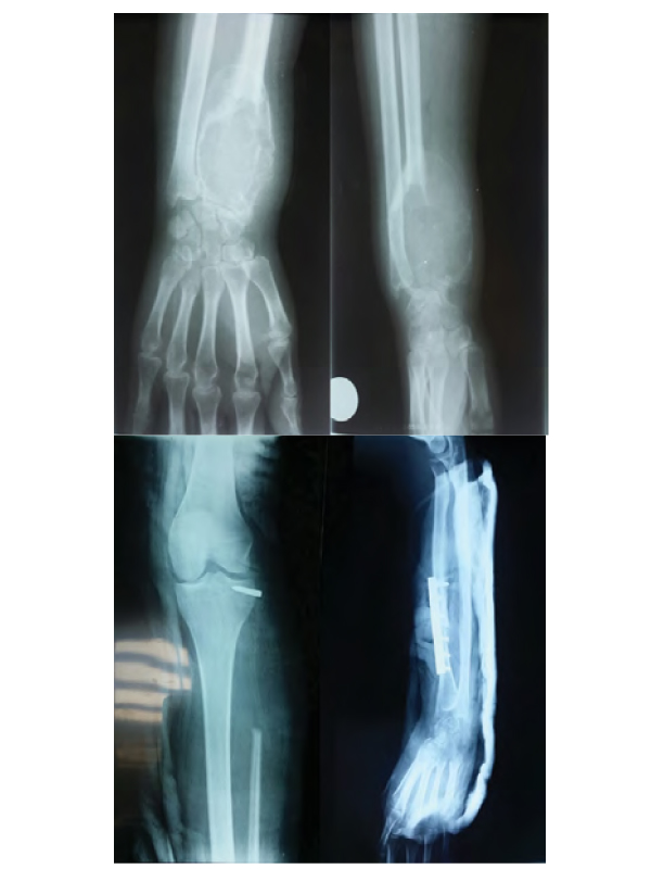
Figure 1: Standard radiography appearance showing an extensive and lytic epiphyso-metaphyseal tumor of the distal radius. Figure 2: Standard radiography aspect showing the resection-reconstruction by a free fibular transfer and osteosynthesis by a screwed plate.
 Awards Nomination