PHONE
+44-7482-878921
+44-7482-878921
2376-0249
Clinical-Medical Image - International Journal of Clinical & Medical Images (2023) Volume 10, Issue 7

Author(s): Zenjali Sara*, Elgraini Soumya, Fikri Meriem, El kettani Najwa, Jiddane Mohamed and Touarsa Firdaous
Department of Neuroradiology, Hôpital Des Spécialités de Rabat, Mohammed V University Hospital, Rabat, Morocco
Received: 07 June 2023, Manuscript No. ijcmi-23-101682; Editor assigned: 08 June 2023, Pre QC No. P-101682; Reviewed: 23 June 2023, QC No. Q-101682; Revised: 28 June 2023, Manuscript No. R-101682; Published: 05 July 2023, DOI:10.4172/2376-0249.1000901
Citation: Sara Z, Soumya E, Meriem F, Najwa EIK, Mohamed J and Firdaous T. (2023) Herpetic Encephalitis: Typical Ascpect. Int J Clin Med Imaging 10: 901.
Copyright: © 2023 Sara Z, et al. This is an open-access article distributed under the terms of the Creative Commons Attribution License, which permits unrestricted use, distribution, and reproduction in any medium, provided the original author and source are credited.
The Herpes Simplex Virus (HSV) is the most prevalent pathogen causing infectious encephalitis, accounting for nearly half of all confirmed cases [1]. This condition is severe, with a case fatality rate of 10-20% and a high incidence of long-term complications [2]. HSV is classified into two types: HSV type 1, which commonly manifests as viral reactivation in adults and HSV type 2, primarily affecting neonates as a primary infection [3]. Our case is the case of a 61 years old patient, without any particular pathological antecedents, who presented with a sudden onset of the following symptoms:
Confusion and alteration of consciousness,intense headache,motor deficit of the 4 limbs, deviation of the face and eyes and mutism.
The clinical examination revealed a GCS of 9, a fever of 39, a stiff neck, a tetraparesis associated with a myocolony.
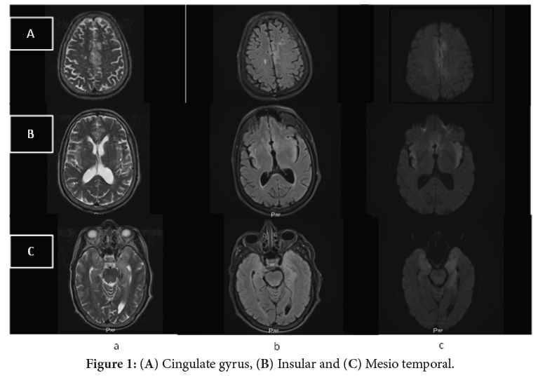
A cerebral CT scan performed first was normal, hence the interest of a cerebral MRI. Axial sections of his brain MRI in T2-weighted (a), Fluid attenuated inversion recovery (Flair)-weighted (b) and Diffusion-weighted (c) sequences showed bilateral asymmetric cortico-subcortical parenchymal damage in hyper-intensities T2-weighted and (FLAIR), with a a restricted diffusion (DWI) in affected area (Figure 1).
Encephalitis caused by the herpes simplex virus (HSV) is characterized by inflammation of the brain tissue, resulting in neurological impairment. It primarily affects the temporal and frontal lobes of the brain, including the limbic system. Typical symptoms encompass headache, changes in consciousness, disorientation or confusion, neurological deficits such as language difficulties (aphasia), muscle pain (myalgia), respiratory or gastrointestinal problems, seizures and fever [1,2].
Early diagnosis and treatment are crucial in this medical emergency. Typically, the diagnosis is confirmed through lumbar puncture and analysis of cerebrospinal fluid using Polymerase Chain Reaction (PCR) testing. While Computed Tomography (CT) scans do not usually reveal any pathological findings, Magnetic Resonance Imaging (MRI) is highly valuable due to its high sensitivity. MRI can detect bilateral asymmetrical signal abnormalities in the temporal lobes, fronto-basal cortex, insula and cingulate, presenting as hyperintensities on T2-weighted and Fluid-Attenuated Inversion Recovery (FLAIR) images. Additionally, Diffusion-Weighted Imaging (DWI) can reveal hyperintense lesions with restricted Apparent Diffusion Coefficient (ADC) in affected areas during the acute phase. Gadolinium-enhanced MRI may also show areas of contrast enhancement. In severe cases, HSVE may present with cortical and subcortical hemorrhagic necrosis, characterized by hypointense T2* signal [1,3].
Herpetic; Encéphalitis; MRI
None of the authors has any conflicts of interests to disclose.
[1] Zhang L, Zhang L, Li F, Liu W, Tai Z and Xu Z, et al. (2023). When herpes simplex virus encephalitis meets antiviral innate immunity. Front Immunol 14: 1118236.
Google Scholar, Crossref, Indexed at
[2] Genet P, Merkler D, Zerlauth JB and Fracasso T. (2023). Incidental discovery of herpes simplex virus encephalitis by post-mortem MRI. Forensic Sci Int 7: 100310.
Google Scholar, Crossref, Indexed at
[3] Bertrand A, Leclercq D, Martinez-Almoyna L, Girard N, Stahl JP and De-Broucker T, et al. (2017). MR imaging of adult acute infectious encephalitis. Med Mal Infect 47: 195-205.
 Awards Nomination