PHONE
+44-7482-878921
+44-7482-878921
2376-0249
Clinical-Medical Image - International Journal of Clinical & Medical Images (2023) Volume 10, Issue 8

Author(s): Abir El Hanzi*, El Mehdi Ait Belhaj, Hatim Essaber, Assad El Bakkari, Soukaina Allioui and Hounayda Jerguigue
Department of Radiology, National Institute of Oncology, Mohamed V University, Rabat, Morocco
Received: 07 August 2023, Manuscript No. ijcmi-23-111217; Editor assigned: 08 August 2023, Pre QC No. P-111217; Reviewed: 22 August 2023, QC No. Q-111217; Revised: 23 August 2023, Manuscript No. R-111217; Published: 30 August 2023, DOI:10.4172/2376-0249.1000908
Citation: Hanzi AE, Belhaj EMA, Essaber H, Bakkari AE and Allioui S, et al. (2023) Heterotopic Ossification in a Midline Laparotomy Scar. Int J Clin Med Imaging 10: 908.
Copyright: © 2023 Hanzi AE, et al. This is an open-access article distributed under the terms of the Creative Commons Attribution License, which permits unrestricted use, distribution and reproduction in any medium, provided the original author and source are credited.
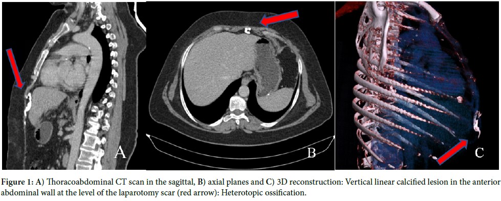
We report a case of a 59-year-old woman who was referred to our radiology department for a thoracoabdominal Computed Tomography (CT) as part of follow-up assessment after a surgical resection of gastrointestinal stromal tumor performed one year previously. The patient presented no complaints. The CT scan of the abdomen showed a 6 cm longitudinal, linear, calcified lesion at the level of the incision scar on the anterior abdominal wall adjacent to the xiphoid region. This finding matched the description of heterotopic ossification, a subtype of myositis ossificans traumatica.
Heterotopic Ossification (HO), also known as myositis ossificans traumatica, is a rare and benign condition that refers to the development of mature lamellar bone at extra-skeletal sites [1]. Patients who underwent a laparotomy rarely experience this rare complication. In a survey of post-operative CT scans, Kim J, et al. discovered that 25% of all patients experiencing open abdominal surgery had HO [2]. Ectopic bone development in midline incision scars usually occurs within a few months of surgery and almost invariably within the first year. In our patient, heterotopic ossification was observed within one year of the surgical procedure [3]. The literature demonstrated a high variability in size; nevertheless, Wang et al. reported that 42% of patients had acquired heterotopic ossification measuring 10 cm in length [1]. In our case, the measurement was 6 cm. Although it is asymptomatic in most cases, it may cause discomfort and chronic abdominal pain. It is crucial to radiologically distinguish this benign entity from other postsurgical complications such as foreign object retained, abdominal wall infection, as well as from primary or metastatic bone tumor [3]. Our patient was asymptomatic and the discovery was incidental during the follow-up. Most patients with heterotopic ossification are asymptomatic and do not require medical treatment. However, if the patient exhibits symptoms, the lesion should be excised [3].
Heterotopic ossification; Computed tomography; Anterior abdominal incision scar
None of the authors have any conflict of interest to disclose.
[1]Ferreira C, Gomes C, Melo A, Tenreiro N and Pinto B, et al. (2017). Heterotopic mesenteric and abdominal wall ossification–two case reports in one institution. Int J Surg Case Rep 37: 22-25.
Google Scholar, Crossref, Indexed at
[2]Kim J, Kim Y, Jeong WK, Song SY and Cho OK. (2008). Heterotopic ossification developing in surgical incisions of the abdomen: Analysis of its incidence and possible factors associated with its development. J Comput Assist Tomogr 32: 872-876.
Google Scholar, Crossref, Indexed at
[3]Kim H. (2018). Heterotopic ossification in the abdominal wall after exploratory laparotomy. J Trauma Inj 31: 177-180.
 Awards Nomination