PHONE
+44-7482-878921
+44-7482-878921
2376-0249
Case Blog - International Journal of Clinical & Medical Images (2014) Volume 1, Issue 1

Author(s): Jiann-Jy Chen
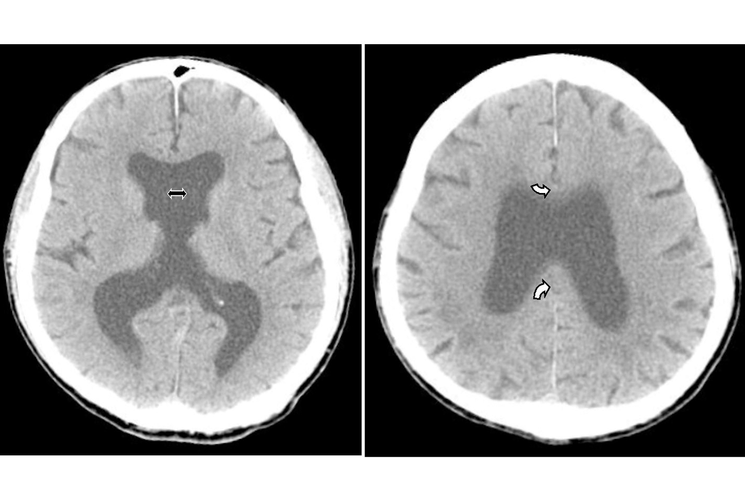
A 79 year-old healthy man, a retired teacher, came to our hospital because he has become forgetful for three years. There was no headache, vertigo, paresthesia, hearing block, ataxia or other neurological signs. He was alert and oriented to time, place and people, but electroencephalography revealed mildly diffuse low background activities so mild diffuse cerebral dysfunction was impressed. Then, computed tomography demonstrated mono-ventricle and fusion of posterior frontal and parietal lobes (Figure) rather than sulci dilatation or subarachnoid space widening. Afterward, conservative treatment and follow-up was recommended.
Holoprosencephaly (HPE), once known as arhinencephaly, is the most common developmental disorder of the human forebrain and involves incomplete separation of the cerebral hemispheres, and selective depletion of a subset of cortical interneurons exists. HPE is estimated to occur in up to one out of 8,000~10,000 live and stillbirths [1,2]. Chromosomal abnormalities account for the most commonly identified cause. According to the degree of cerebral hemispheric separation, HPE is classified into alobar, semilobar, lobar and middle interhemispheric variant types. The middle interhemispheric variant type of HPE was first described in 1993 [3]. It is also referred to as “syntelencephaly, representing that the posterior frontal lobe and the parietal lobe are not properly separated, but the rostrobasal forebrain properly separates [4], as in our case patient.
Common medical problems of HPE in children include hydrocephalus, seizures/epilepsy, motor impairment, pulmonary diseases, gastrointestinal problems, oromotor dysfunction, feeding and nutrition, endocrine dysfunction, hypothalamic dysfunction, ophthalmologic problems, and craniofacial-related complications. Therefore, mortality is high in newborns with HPE, and only a minority of the sufferers could survive beyond the neonatal period [1]. Our case patient is indeed a rare curiosity who has non-syndromically survived into adulthood and even aged-hood. Herein, we report the case for neurologic and neuroradiological interest.
Reference
Corresponding author
Jiann-Jy Chen
Department of Neurology
China Medical University Hospital
Taichung
Taiwan
 Awards Nomination