PHONE
+44-7482-878921
+44-7482-878921
2376-0249
Clinical-Medical Image - International Journal of Clinical & Medical Images (2023) Volume 10, Issue 1

Author(s): Diallo Ibrahima Dokal1*, Traore Mohamed Wend-Yam2, El Ouali Ibtissam2, Zahi Hiba2, Moatassim Nabil Billah2 and Nassar Iitimad2
1Department of Imaging, Children hospital, Mohammed V university, Rabat, Morocco
2Department of Imaging, Central Radiology, Mohammed V university, Rabat, Morocco
Date of Submission: 20 January 2023, Manuscript No. ijcmi-23-87480; Editor assigned: 21 January 2023, Pre QC No. P-87480; Reviewed: 26 January 2023, QC No. Q-87480; Revised: 29 January 2023, Manuscript No. R-87480; Published: 05 February 2023, DOI:10.4172/2376-0249.1000871
Citation: Dokal ID, Wend-YamTM, Ibtissam EO, Hiba Z and Billah MN, et al. (2023) Hypophysive Hemochromatosis: A Rare Cause of Hypothyroidism. Int J Clin Med Imaging 10:871.
Copyright: © 2023 Dokal DI, et al. This is an open-access article distributed under the terms of the Creative Commons Attribution License, which permits unrestricted use, distribution, and reproduction in any medium, provided the original author and source are credited.
Pituitary hemochromatosis is an accumulation of excess iron in the anterior lobe of the pituitary gland. It occurs in patients receiving repeated transfusions. The discovery of this disease is usually due to endocrine disorders: diabetes mellitus, hypogonadal hypo-gonadotropism. In rare cases a hypothyroidism can reveal it as in the following cases.
Hemochromatosis refers to excess iron build up. This build-up can occur at various sites, including the pituitary gland. Diabetes mellitus and hypogonadotropic hypogonadism are the two main manifestations of this condition. In rare cases, uncommon manifestations such as hypothyroidism can reveal it. We illustrate this type of manifestation through the case below.
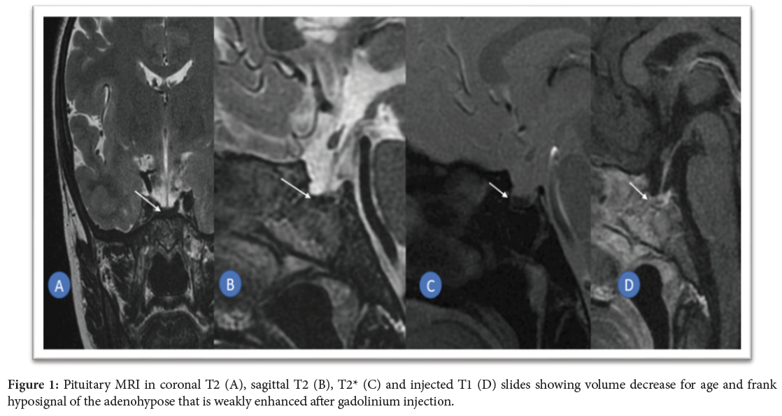
A 25-year-old patient, under surveillance for beta-thalassemia since the first month of life, was referred to our facility for investigation of hypothyroidism. His history showed repeated transfusions, while the biological workup showed elevated ferritin levels in contrast to a decrease in TSH and peripheral hormones (LT3, LT4). A pituitary MRI was performed (Figure 1).
Iron overload, known as hemochromatosis, occurs in special circumstances requiring repeated transfusions (hemophilia, beta-thalassemia, sickle cell disease). It can affect the liver, pancreas, thyroid, parathyroid and pituitary glands, thus altering the function of these organs. Diabetes mellitus is one of the common manifestations of this disease [1].
The pituitary damage linked to hemochromatosis is far from being unknown; it only involves the anterior lobe of the pituitary gland: the adenophyseal gland, whose role is essentially endocrine. Gonadotropic cells are preferentially those affected. The accumulation of iron in the adenohypophysis leads to a deficiency of anteropituitary hormones (TSH, FSH, LH), explaining the hypogonadism and on rare occasions the adrenal deficiency or hypothyroidism occurring in patients with pituitary hemochromatosis [1]. Magnetic resonance imaging to the rescue of biological work up plays a fundamental role in the diagnosis of this endocrinopathy. It shows a significant drop in the signal of the anterior lobe of the pituitary gland, due to the decrease in T2 relaxation time and to the inhomogeneity of the magnetic field resulting from the intracellular accumulation of iron. The T2* weighted sequence [2], is the most sensitive to identify this build-up, due to the magnetic susceptibility, and allows the demonstration of hemosiderin deposits in the form of a frank hyposignal that correlates with the amount of iron. However, T2* images may be artifacted at the base of the skull (sella turcica, sphenoid), due to this same magnetic susceptibility .Moreover, pituitary overload is accompanied by a decrease in the volume of the adeno-pituitary gland with weak enhancement after injection [3]. This typical aspect of the imaging was the one found in our patient, which allowed us to retain the diagnosis of this condition after confrontation with the clinicobiological findings.
Hemochromatosis is a condition occurring in patients receiving iterative transfusions. It can affect various endocrine glands including the pituitary gland. MRI through T2 and T2* sequences come to the rescue of the biological workup to support the diagnosis. Its discovery should lead to a search for other localizations of hemochromatosis.
Hemochromatosis; Pituitary gland; Transfusions
I would like to express my gratitude to my professors and all the colleagues who participated in the completion of this work.
This research received no specific grant from any funding agency in the public, commercial, or not for profit sectors.
The authors declare no conflict of interest.
[1] Khelifi D., Debbabi W., Soomauroo S., Kharrat I. and Samet, et al. (2020). Pituitary hemochromatosis: About 3 cases. J Belg Soc Radiol 24: 296.
Google Scholar, Crossref, Indexed at
[2] Sparacia G., Iaia A., Banco A., D'Angelo P. and Lagalla R, et al. (2000). Transfusional hemochromatosis: Quantitative relation of MR imaging pituitary signal intensity reduction to hypogonadotropic hypogonadism. Radiology 215: 818-823.
Google Scholar, Crossref, Indexed at
[3] Noetzli LJ, Panigrahy A., Mittelman SD., Hyderi A., Dongelyan A., et al. (2012). Pituitary iron and volume predict hypogonadism in transfusional iron overload. Am J Hematol 87: 167-171.
 Awards Nomination