PHONE
+44-7482-878921
+44-7482-878921
2376-0249
Clinical Image - International Journal of Clinical & Medical Images (2014) Volume 1, Issue 5

Author(s): Ayman Karkar
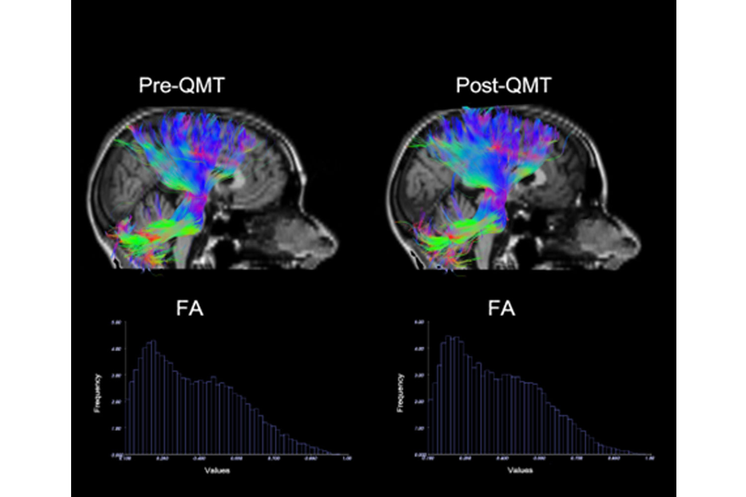
Maintaining neuroplasticity is an important goal that can be stimulated through training. The Quadrato Motor Training (QMT) is a new sensorimotor training which has recently been reported to increase functional connectivity and cerebellar activity. QMT was further found to improve cognitive functions, including creativity, spatial cognition and reading. Here we report the effects of a pilot diffusion tensor imaging (DTI) tractography study related to the effects of 12-weeks of daily QMT. White matter (WM) changes were evaluated from a healthy female volunteer, using Fractional Anisotropy (FA) values computed from DTI [1,2]. We found post-QMT FA increments of a complex WM projection system, including the cerebellar and corona radiata tracts, in comparison to pre-QMT (total fibers volume form 135745 mm3 to 140611 mm3; total number of fibers from 2809 to 3272). This complex projection system is closely related to sensorimotor and higher cognitive functions [3,4]. Although preliminary in their nature, the present exploratory work strengthens previous findings related to the effects of sensorimotor training. More specifically, the cerebellar reorganization support previous results regarding the effects of QMT [5] and may point to a causal relationship between sensorimotor training and plasticity. Pre- and post-QMT (left and right columns, respectively) DTI fiber tracts superimposed on T1-Weighted MR image. The color-coded indicates the local mean direction of the tracks: red, left–right; green, anterior–posterior; blue, superior–inferior. Post-QMT FA values of the cerebellar and of the corona radiata tracts show an higher frequency in comparison to pre-QMT, as illustrated in the two diagrams.
*Corresponding author: Ayman Karkar, Department of Nephrology, Kanoo Kidney Center, Dammam Medical Complex, PO Box 11825, Dammam 31463, Kingdom of Saudi Arabia, Tel: 00966-3-815- 5740; Fax: 00966-3-815-5625; E-Mail:aymankarkar@yahoo.com
 Awards Nomination