PHONE
+44-7482-878921
+44-7482-878921
2376-0249
Clinical-Medical Image - International Journal of Clinical & Medical Images (2023) Volume 10, Issue 6

Author(s): Abir Lemrabet*, Yassine Zerhari, Asaad El Bakkari, Soukaina Allioui, Hatim Essaber, Hounayda Jerguigue, Youssef Omor and Rachida Latib
Department of Radiology, National Institute of Oncology of Rabat, Morocco
*Corresponding Author:
Abir Lemrabet
Department of Radiology
National Institute of Oncology of Rabat, Morocco
Tel: +212 653828900
E-mail: lemrabetabir20@gmail.com
Received: 12 May 2023, Manuscript No. ijcmi-23-98579; Editor assigned: 15 May 2023, Pre QC No. P-98579; Reviewed: 29 May 2023, QC No. Q-98579; Revised: 05 June 2023, Manuscript No. R-98579; Published: 12 June 2023, DOI: 10.4172/2376-0249.1000897
Citation: Lemrabet A, Zerhari Y, Bakkari ELA, Allioui S, Essaber H and Jerguigue H, et al. (2023). Interruption of VCI with Azygos Continuation. Int J Clin Med Imaging 10: 897.
Copyright: © 2023 Lemrabet A, et al. This is an open-access article distributed under the terms of the Creative Commons Attribution License, which permits unrestricted use, distribution, and reproduction in any medium, provided the original author and source are credited.
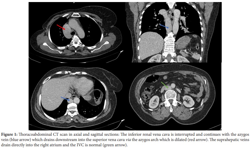
A 40-year-old female patient, with no previous cardiovascular history, underwent a thoracic-abdominopelvic CT scan as part of the extension assessment of a ductal carcinoma in situ of the breast, incidentally showing an interruption of the IVC with azygos continuation (Figure 1).
Azygos continuation of the inferior vena cava is also known as absence of the hepatic segment of the IVC with azygos continuation. It is a rare vascular anomaly especially when not associated with congenital heart disease, asplenia or polysplenia syndromes. During embryonic life, the IVC is formed of 4 segments (hepatic, prerenal, renal and postrenal). The interruption of the IVC most likely results from a failure of fusion of the embryological yolk and subcardinal portions of the IVC. Therefore, azygos drainage occurs downstream into the superior vena cava via the enlarged azygos arch. The suprahepatic veins drain directly into the right atrium. This congenital anomaly does not have any specific presenting symptoms and its diagnosis is evoked on frontal chest radiography (dilatation of the azygos with outward displacement of the right paravertebral line) or on ultrasound (absence of visualization of the suprarenal vena cava). CT scan and contrast-enhanced phlebography may be helpful.
Recognition of this vascular anomaly preoperatively may be important in planning extracorporeal circulation and avoiding difficulties in cardiac catheterization [1-3].
Continuation azygos; Veine cave inférieure; Tomodensitométrie
None of the authors has any conflicts of interests to disclose.
[1] Bass JE, Redwine MD, Kramer LA, Huynh PT and Harris Jr JH. (2000). Spectrum of congenital anomalies of the inferior vena cava: Cross-sectional imaging findings 1: (CME available in print version and on RSNA Link). Radiographics 20: 639-652.
Google Scholar, Crossref, Indexed at
[2] Ghandour A, Partovi S, Karuppasamy K and Rajiah P. (2016). Congenital anomalies of the IVC—embryological perspective and clinical relevance. Cardiovasc Diagn Ther 6: 482.
Google Scholar, Crossref, Indexed at
[3] Saito T, Watanabe M, Kojima T, Matsumura T and Fujita H, et al. (2011). Successful blood sampling through azygos continuation with interrupted inferior vena cava a case report and review of the literature. Int Heart J 52: 327-330.
 Awards Nomination