PHONE
+44-7482-878921
+44-7482-878921
2376-0249
Case Blog - International Journal of Clinical & Medical Images (2016) Volume 3, Issue 11

Author(s): Patrick Francois Tarquino, John Elkin Pedraza and Juan S Barajas-Gamboa
Introduction: A 69 year-old female was incidentally diagnosed to have an intra-hepatic atherosclerosis disease on 3D Reconstruction of Abdominal Computed Tomography Angiography performed at the Emergency Room, while surgeons and urologist were evaluating an acute abdominal pain in a patient with several comorbidities. The radiological findings and differential diagnosed are briefly discussed. For academic, research and publication purposes, the patient signed an informed consent
Keywords: Abdominal pain; Atherosclerosis disease; Intrahepatic; Vascular biology.
Clinical Scenario/Question: A previously well-known 69 year-old female with a large medical history of Systemic Lupus Erythematous (SLE), Lupus Nephritis (LN), Hypertension (High Blood Pressure), Hypothyroidism, Antiphospholipid Syndrome (APS) Chronic renal disease stage III B, episode of extensive Deep Vein Thrombosis (DVT) in the right lower limb andPulmonary Embolism (PE), an episode of Multiple Organ Failure (MOF), that required Renal Replacement Therapy (RRT), was brought to the emergency room consulting for a suddenly acute abdominal pain focused in the right lumbar region and right flank, intensity 8/10 in the Visual Analog Scale (VAS), associated with nauseas, vomiting.
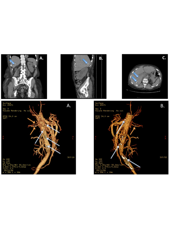
Abdominal CT scan and CT urography were performed by general surgeon recommendation. Abdominal CT scan images showed a hypodense, homogeneous, well- demarcated lesion of 42 × 38 mm, without the presence of enhancement, nearly the bifurcation of the right common iliac artery, which implies adnexal origin. No free abdominal fluid was observed and no intra-abdominal collections were considered (Figure 1).
CT urography reported right hydroureteronephrosis, hepatic residual calcifications and pelvic cystic lesion with no relevant anatomical impact in this area. Secondary to the wide range of findings and unclear principal diagnosis, a 3D Reconstruction of Abdominal Computed Tomography Angiography was ordered, incidentally diagnosed to have an intra-hepatic atherosclerosis (Figure 2). What is the possible diagnosis?
1. Intra-hhepatic atherosclerosis
2. Hematogenous metastasis
3. Giant cell arteritis
4. Hemangiomas
5. Polyarteritis nodosa
The atherosclerosis disease is a common disorder in general population characterized by the present of endothelial damage, secondary to the deposition in plaques of fatty material on their inner walls, silently and slowly blocking the lumen of the arteries, putting the blood flows at risk.
This medical situation may finally develop ischemic and embolic event [1,2]. This pathophysiologic disorder may be affected or exacerbated by other comorbidities in some patients such as exogenous and endogenous factors. The atherosclerosis disease is commonly found in vessels of great or middle caliber, most of them illustrated to have a rich laminar blood flow, which allow the development of atherosclerotic plaques [3,4]. In this medical case, the patient presented an important autoimmune background; including SLE, APS, a prothrombotic predisposition, calcium, lipids and inflammation factors deposits, that may lead a deleterious role in the genesis of atherosclerosis in minor vessels that are not expected to be affected. Finally, the blood flow compromised in these vessels can be reflected in lesions of important organs such as kidneys, brain, heart and splanchnic bed [5,6,7].
In conclusion, Intra-Hepatic Atherosclerosis Disease discovered at the emergency room in the clinical scenario of the acute abdominal pain, has to be considered by clinicians and emergency physicians as differential diagnosis.
 Awards Nomination