PHONE
+44-7482-878921
+44-7482-878921
2376-0249
Case Blog - International Journal of Clinical & Medical Images (2016) Volume 3, Issue 4

Author(s): Madhuri Rajesh Chandnani
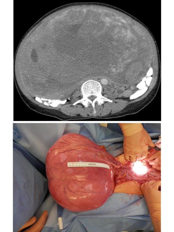
A 60 year old female with no significant past medical history presented to the Emergency department for worsening abdominal distension of 3 years duration Review of systems positive only for a weight loss of about 10 lbs in last 7 months, and negative for all others including any GI or GU symptoms. CT abdomen and pelvis with contrast demonstrated a very large mass measuring 27 × 15 × 25 cm3 , possibly arising from the uterus or ovaries (Figures 1-4). Also, seen were enlarged left renal and pelvic varices due to compression of the left renal vein and IVC by the mass. CT chest significant for possible old granulomatous disease an exploratory laparotomy was performed and revealed an enlarged uterus extending up to the xiphoid.
All the other abdominal and pelvic structures appeared normal. A total abdominal hysterectomy and bilateral salpingo-oophorectomy was performed. The uterus was 37.5 × 22.5 × 18.5 cm3 , with multiple intramural masses with a maximum diameter up to 14.5 × 14.5 × 10.5 cm3 . Pathology revealed leiomyosarcoma with intrauterine satellite nodules, confined to the uterus; otherwise benign parametrium with benign ovaries, fallopian tubes and cervix. Leiomyosarcoma of the uterus is an extremely rare, but highly aggressive uterine malignancy with high risk of recurrence and death [1]. Surgery with post-operative surveillance is the preferred treatment in patients with disease confined to pelvis, and in patients with symptoms like pain or vaginal bleeding. Adjuvant chemotherapy is recommended in patients with intra-abdominal spread or distant metastases [2]. Our patient here follows with Gynecologic oncology for postoperative surveillance and has been asymptomatic one month since the surgery.
 Awards Nomination