PHONE
+44-7482-878921
+44-7482-878921
2376-0249
Clinical-Medical Image - International Journal of Clinical & Medical Images (2021) Volume 8, Issue 2

Author(s): Tripti Ramya* and Santoshi
Clinical Image
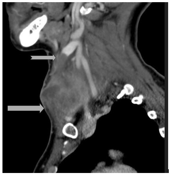
A 44-year-old female with PMH of Hepatitis C, seizures, bipolar disease, cervical cancer s/p hysterectomy and IV drug abuse presented to the ED with a neck mass of roughly 2 weeks duration along with constant “achy” pain of 7/10 severity radiating to left side of her back and shoulder. During the 7 days prior to her ED visit, the mass was increasing in size and causing difficulty breathing and swallowing. Old records revealed that the patient presented to the ED 2 weeks prior, with high fever, lethargy and oral abscess and was on treatment with oral Clindamycin. No neck swelling was visible at that point and she was treated with IV Metronidazole and Amoxicillin/Clavulanate. Initial physical examination showed a 6 × 5 cm solitary, tense, tender, erythematous swelling in zone 2 on the left side of neck, which did not move with deglutition. Her WBC count was normal. A CT scan of the soft tissue of the neck showed a loculated left-sided neck abscess measuring 6.6 × 6.0 × 4.0 cm and a large thrombus in the anterior branch of IJV. A Chest X-ray ruled out major pulmonary complications. Laboratory results revealed a negative blood culture and wound culture positive for multi-drug resistant strains of E. coli and S. aureus. The patient was managed with airway monitoring, IV Ampicillin/Sulbactam and drainage of the abscess. Unfortunately, after being explained the risks and consequences, she left against medical advice on the 3rd day of hospitalization and was lost to follow-up (Figure 1). Lemierre’s syndrome is suppurative thrombophlebitis of the Jugular vein frequently preceded by oropharyngeal infection [1]. The overall incidence is 3.6 in one million people [2] and mortality rate is up to 17% [3]. The most common organisms involved are oral anaerobes like Fusobacterium species [4]. Skin flora and nosocomial pathogens are common in the background of central line placement or IV drug use. Patients usually present with fever and neck pain. Preferred methods of diagnoses are cultures from the site and Contrast-enhanced CT scan of neck that demonstrates filling venous defect [5]. Common complications are metastases of septic emboli to lungs and other organs. Carotid sheath involvement may also lead to carotid artery mycotic aneurysms and rupture; hoarseness; Horner’s syndrome. The important principles of treatment are IV antibiotics and surgical intervention. It is crucial to cover oral anaerobes with Ampicillin-Sulbactam, Piperacillin-Tazobactam, Ticarcillin-Clavulanate or single Carbapenem for at least four weeks, including a minimum of two weeks intravenous therapy followed by oral therapy [2]. Vancomycin can be added for patients with MRSA positive risk factors. Surgical intervention/endovascular treatment is recommended in the setting of sepsis or when no response to antibiotics is noted [6,7] like in our patient above. Anticoagulation is controversial, recommended with thrombus extension and sepsis. It was not started in this case due to absence of prior imaging for comparison and no septic findings. To summarize, management of Lemierre’s syndrome involves treatment of sources of infection, covering oral anaerobes/skin flora persusceptibility and monitoring for complications.
Keywords: Tetragon; Caudal regression syndrome
 Awards Nomination