PHONE
+44-7482-878921
+44-7482-878921
2376-0249
Clinical-Medical Image - International Journal of Clinical & Medical Images (2021) Volume 8, Issue 5

Author(s): Khaoula Sibbou, Olaia Chalh, Meryem Fikri, Mohamed Jiddane, Firdaous Touarsa
Clinical Image
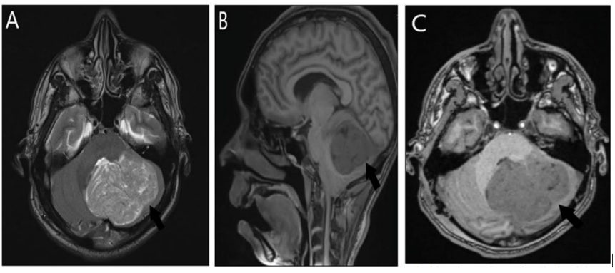
Lhermitte-Duclos disease, also called dysplastic cerebellar gangliocytoma, manifests in young individuals. Most patients’ symptoms stem from increased intracranial pressure and hydrocephalus. A slowly progressive cerebellar syndrome, megalocephaly and mental retardation are less common clinical features. It is considered a hamartomatous lesion, often associated with Cowden’s disease. On a CT examination, dysplastic cerebellar gangliocytoma presents as a low-density cerebellar mass, which may contain calcification and does not show enhancement. At MRI, it reveals most frequently as a left hemispheric cerebellar mass with folial or “tiger-striped” pattern (Figure 1). This tumor is typically iso-and hypointense on T1-weighted images and hyperintense on T2WI with characteristic alternating bands of different signal intensity relative to gray matter. Most cases demonstrate little or no enhancement, although patchy enhancement of the tumor has been described in some series probably related to the deep running veins between the folia seen on SWI. Treatment of choice is surgery. In symptomatic patients it may be necessary to place a ventricular shunt catheter to treat hydrocephalus.
Keywords: Lhermitte-Duclos; MRI; CT
 Awards Nomination