PHONE
+44-7482-878921
+44-7482-878921
2376-0249
Clinical Image - International Journal of Clinical & Medical Images (2014) Volume 1, Issue 3

Author(s): Int J Clin Med Imaging 2014
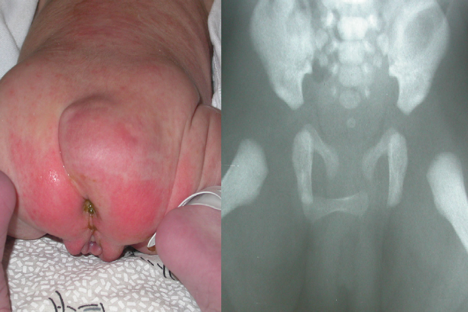
A female infant, born at 39 weeks by caesarian section, had a large mass on the right buttock. Physical examination and the baby’s general well-being were otherwise normal. Plain film revealed an “accessory” bone horizontally oriented and superimposed on the right hemi pelvis (arrowheads) whose location corresponded with the mass. In fact, this same bone was easily palpable by physical examination. In view of the clinical and radiographic findings there is no doubt that this represents a teratoma. The presence of bony structures in a sacrococcygeal tumor is almost pathognomonic for mature teratoma. In order to determine the full extent of the mass prior to surgery ultrasound and MRI were performed. Both imaging studies showed a retrosacral lesion containing solid and cystic components with a small precoccygeal extension. At age 1 week the mass was surgically resected. The post-operative course was uneventful. Histopathologic evaluation confirmed the preoperative diagnosis of benign mature teratoma. Sacrococcygeal teratoma is a rare congenital tumor arising form pluripotential cells. The incidence is 1 in 40,000 live births. Benign mature teratoma is the most common type. The tumor may contain elements from glia, choroid plexus structures, teeth, bronchial mucosa, skin appendages, muscle and bone. When the mass contains bone it is often similar to a metacarpal or digital bone. Treatment for mature teratoma is surgical resection. Prognosis is good but the prevalence of malignant cells increases with the child’s age. Therefore early diagnosis and resection is of utmost importance. This case is a classic example of a bone-forming mature sacrococcygeal teratoma, diagnosed by plain x-ray.
 Awards Nomination