PHONE
+44-7482-878921
+44-7482-878921
2376-0249
Case Blog - International Journal of Clinical & Medical Images (2016) Volume 3, Issue 5

Author(s): Mahesh Shivaji Chavan
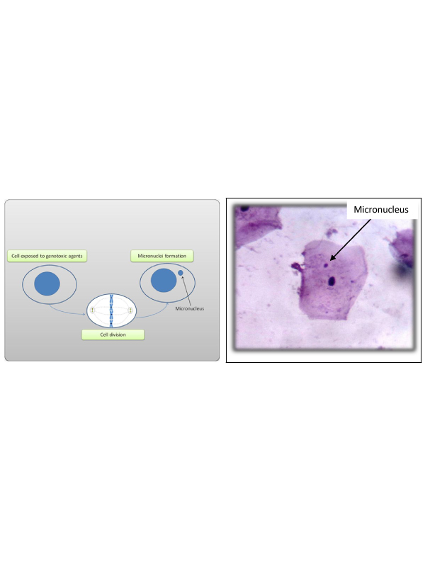
Micronucleus (MN) is a small additional nucleus and is easily identified by light microscopy. Micronucleus scoring could be used as a bio-marker to identify different pre-neoplastic conditions much earlier than the presentation of clinical features and might specifically be exploited in the screening of high-risk population for a specific cancer [1] and it has clearly revealed that the level of baseline chromosome damage in untreated cancer patients is much more than in cancer-free controls. As a result, the prevalence of MN in epithelial cells has been considered a potential tissue-specific indicator of cancer risk [2]. The suggested criteria for identifying micronucleus developed by Tolbert et al. [3] are,
1. Rounded smooth perimeter suggestive of a membrane.
2. Less than a third the diameter of associated nucleus, but large enough to discern shape and color.
3. Staining intensity similar to nucleus.
4. Texture similar to nucleus.
5. Same focal plane as nucleus.
6. Absence of overlap with bridge to nucleus.
In our study we have assessed biomonitoring of DNA damage (in the form of micronuclei) (Figure 1) of oral mucosa cells in individuals exposed to full mouth IOPA survey, panoramic dental radiography and CT scan for maxilla and mandible. Pre and post exposure (after 10 Days) assessment of micronuclei was done. There was statistically significant increase in the number of micronuclei in post exposure to CT scan than pre exposure to CT scan group. This study data indicate that CT scan may induce chromosomal damage to oral epithelial cells. (Figure 2) shows micronucleus in the exfoliated oral epithelial cell 10 days after exposed to CT scan.
 Awards Nomination