PHONE
+44-7482-878921
+44-7482-878921
2376-0249
Clinical-Medical Image - International Journal of Clinical & Medical Images (2023) Volume 10, Issue 9

Author(s): Chaimae Abourak*, Siham Oukassem, Asmae Guennouni, Soukaina Bahha, Amal Lahfidi, Najoua Ech-cherif El Kettani, Meriem Fikri, Mohamed Jiddane and Firdaous Touarsa
Department of Neuro-Radiology, Specialty Hospital, CHU Ibn Sina, Faculty of Medicine and Pharmacy of Rabat, Mohamed V University, Rabat, Morocco
*Corresponding Author:
Chaimae Abourak
Department of Neuro-Radiology
Specialty Hospital, CHU Ibn Sina
Faculty of Medicine and Pharmacy of Rabat, Mohamed V University,
Rabat, Morocco
Tel: +212 673331162
E-mail: chaymaabourak@gmail.com
Received: 11 September 2023, Manuscript No. ijcmi-23-113512; Editor assigned: 12 September 2023, Pre QC No. P-113512; Reviewed: 20 September 2023, QC No. Q-113512; Revised: 21 September 2023, Manuscript No. R-113512; Published: 28 September 2023, DOI:10.4172/2376-0249.1000912
Citation: Abourak C, Oukassem S, Guennouni A, Bahha S and Lahfidi A, et al. (2023). Multi-Cystic Parotids. Int J Clin Med Imaging 10: 912.
Copyright: © 2023 Abourak C, et al. This is an open-access article distributed under the terms of the Creative Commons Attribution License, which permits unrestricted use, distribution and reproduction in any medium, provided the original author and source are credited.
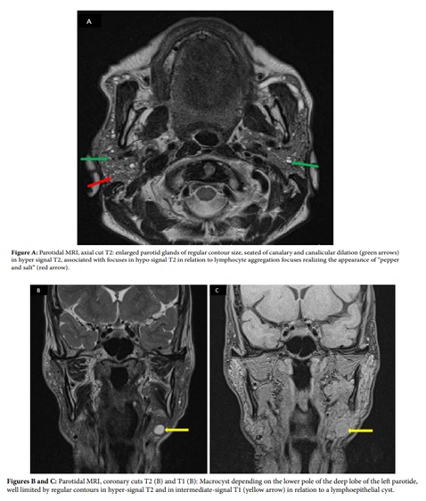
A 68-year-old woman, type II diabetic for five years on oral antidiabetics, has had a recurring left parotid tumor for 10 years associated with dry mouth and eyes. The clinical examination reveals a deep paroid mass that is well limited, not painful at palpation and without other inflammatory signs in sight. The rest of the clinic examination is unparalleled. The ultrasound showed multiple bilateral parotid microcysts with canal dilation. A parotid MRI was performed (Figures A, B and C). The diagnosis of primary Gougerot-Sjögren syndrome revealed by multi-cystic parotides was accepted. Sjogren syndrome is a chronic autoimmune disease characterized by the inflammation and destruction of the exocrine glands, mainly the tear and salivary glands, resulting in a loss of tear and saliva production [1,2]. The syndrome is considered primary when it occurs in the absence of other autoimmune diseases, such as rheumatoid arthritis, systemic lupus erythematosus, polymyositis and multiple sclerosis [1-3]. According to a study of the autopsy reports, Hudson estimated that the incidence of Sjögren syndrome was 1 case per 255 inhabitants [3]. Diagnostic criteria have constantly changed over the years and controversy over the most accurate criteria persists [2]. Although the most typical symptoms of Sjögren’s syndrome are dry eyes and mouth, clinical presentations are diverse [2]. Most patients with primary Sjögren syndrome suffer from diffuse hypertrophy of the salivary glands, mainly the parotid glands. Clinically, there may be episodic, recurring, or permanent unilateral or bilateral swelling [1-3]. There are several imaging methods to explore the salivary glands, especially the parotid glands. Mainly ultrasound, Tomodensitometry (TDM), Magnetic Resonance Imaging (MRI) and sialography. However, parotid MRI remains the test of choice for exploring parotic glands [2]. The MRI shows multiple mixed focuses in hyposignal and hypersignal in T2, realizing the “pepper and salt” appearance that can be considered as evoking Gougerot-Sjogren syndrome. These hyper-intensive focuses in T2 may correspond to the dilated tubular system (sialectasia), while those hypo-intensive in T2 may be due to focuses of lymphocyte aggregation [1-3]. Our case showed multiple bilateral microcysts with a single lower left polar macrocyst in hypersignal T2 and intermediate signal T1, suggesting benign lymphoepithelial cysts in the background of Gougerot-Sjogren syndrome. In addition, these cysts are associated with multiple mixed focuses in hyper- and hypo-signal T2, realizing the “pepper and salt” aspect and evoking the Gougerot-Sjogren syndrome. Multi-cystic parotid affliction is rare and yet the presence of multiple bilateral parotic cysts is more rare in the context of Gougerot-Sjogren syndrome; only a few cases of this kind have been reported [2]. The diagnosis of Gougerot-Sjogren syndrome should be included in the differential diagnosis for cystic and bilateral parotidal lesions [2]. These main radiological differential diagnoses are HIV-related benign lymphoepithelial lesions, branchial cysts and Warthin’s cystic tumor [1-3]. Histological confirmation is crucial in the diagnosis of Gougerot-Sjogren syndrome by salivary gland biopsy [3]. Patients with this syndrome have a high risk of developing lymphoma. Regular follow-up with these patients is essential for the early detection of lymphoma-inducing signs [1,3].
Parotid; Cyst; Sjogren’s syndrome
None of the authors has any conflicts of interests to disclose.
[1]Plaza G, Dominguez MP and Bueno A. (2003). Bilateral parotid cysts as presentation of Sjögren’s syndrome. J Laryngol Otol 117: 151-152
Google Scholar, Crossref, Indexed at
[2]Seo BF, Ju RK, Kwok, SK and Oh DY. (2014). Unusual Sjögren's syndrome with bilateral parotid cysts. Arch Craniofac Surg 15: 98.
Google Scholar, Crossref, Indexed at
[3]Toh AS, Broomfield SJ, Teh LS, Aslam MB and Duncan G, et al. (2011). Bilateral multicystic parotid masses in primary Sjögren syndrome. Ear Nose Throat J 90: E20-E22.
 Awards Nomination