PHONE
+44-7482-878921
+44-7482-878921
2376-0249
Clinical-Medical Image - International Journal of Clinical & Medical Images (2022) Volume 9, Issue 9

Author(s): Pablo Cisneros Arias*
Department of Ophtalmology, Hospital Clinico Universitario Lozano Blesa, Zaragoza, Spain
Received: 22 August, 2022, Manuscript No. ijcmi-22-73386; Editor assigned: 26 August, 2022, 2022, PreQC No. P-73386; Reviewed: 16 September, 2022, QC No. Q-73386; Revised: 23 September, 2022, Manuscript No. R-73386; Published: 30 September, 2022, DOI: 10.4172/2376-0249.1000847
Citation: Arias PC. (2022) Myelinated Retinal Nerve Fiber Layer (MNFL). Int J Clin Med Imaging 9:847.
Copyright: © 2022 Arias PC. This is an open-access article distributed under the terms of the Creative Commons Attribution License, which permits unrestricted use, distribution, and reproduction in any medium, provided the original author and source are credited.
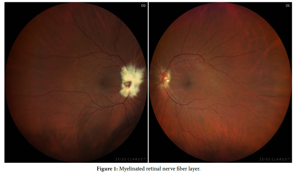
Myelinated retinal nerve fiber layer (MNFL) is a rare congenital anomaly that can appear as opaque greyish-white striated patches on the superficial retina with soft edges that obscure retinal details. It is usually a unilateral condition. Most of the cases are found in the disc. It remains stable over time and has no impact on visual development. Although, on occasions, it is associated with other pathological conditions such as myopia, amblyopia or strabismus, which can darken the visual prognosis. We present a case of a 45-year-old man who attended a routine ophthalmological evaluation after starting treatment with hydroxychloroquine for rheumatic condition. Fundus examination showed papillary myelin fibers in both eyes, predominantly in the right eye. The rest of the examination, such as OCT and visual field tests were unremarkable (Figure 1).
Congenital anomalies ophthalmology retina myelin
 Awards Nomination