PHONE
+44-7482-878921
+44-7482-878921
2376-0249
Case Blog - International Journal of Clinical & Medical Images (2014) Volume 1, Issue 3

Author(s): Int J Clin Med Imaging 2014
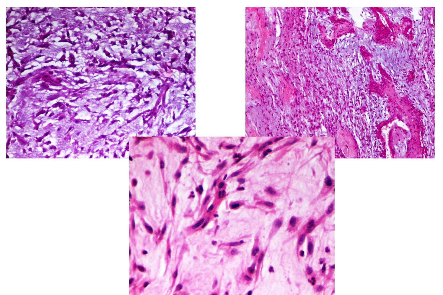
Myxoid leiomyosarcoma of the uterus is an aggressive, recurrent, metastasizing and outstandingly rare form of leiomyosarcoma with an incidence of 0.3-0.67% per 100.000 women. This neoplasm poses diagnostic challenge owing to its rarity and lack of mitotic activity which is a typical criterion for malignancy [1,2]. King et al. [3] first described ‘myxoid leiomyosarcoma of the uterus” in 1982, when they reported on six cases. This tumor arises from the smooth muscle cells of the myometrium or possibly from the blood vessels [2], or it may arise in association with a benign leiomyomatous lesion [1]. Microscopically, the tumor has a remarkable myxoid appearance and displays a highly malignant invasive behavior despite its low mitotic index. Akin to the other described cases of myxoid leiomyosarcoma, bipolar spindle cells without atypia and having a low mitotic index [0-2 in 10 high power fields], and few multinucleated cells are seen in a background of myxoid material. Focally bundles of smooth muscle cells are noticed with areas of necrosis and hemorrhage. Therefore myxoid leiomyosarcoma is an exception to the general rule that mitotic count is determinant to the malignant behavior of smooth muscle tumors. Although it is difficult to establish the nature of the neoplastic cells in the myxoid areas, both light microscopic and immuno-histochemical characteristics shows features of smooth muscle cells in some of the cellular areas [1,2]. The diagnosis should be based on gross and microscopic features and on the aggressive behavior rather than on mitotic index or atypia of the tumor cells [Figure 1-4].
 Awards Nomination