PHONE
+44-7482-878921
+44-7482-878921
2376-0249
Case Blog - International Journal of Clinical & Medical Images (2016) Volume 3, Issue 1

Author(s): Chin-Chung Lin, Wei-Kung Chen and Chun-Hung Chen*
Figure 1: Chest radiograph showing an occupied lesion over the trachea.
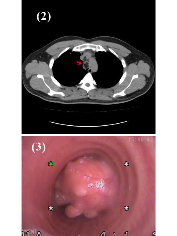
Figure 2: CT of the chest revealed a near obstructing tracheal tumor with low attenuation suggesting a fat containing lesion.
Figure 3: Bronchoscopic image showing a huge rounded tumor with almost 90% lumen obstruction. in the trachea.
Clinical Presentation A 32-year-old man was evaluated for a 6-month history of dyspnea, and non-productive cough. On physical examination, he was found to have stridor breath sounds. A chest radiograph showed a mass over the trachea (Figure 1). A contrast enhanced chest CT showed a low attenuation mass in the trachea (Figure 2). Fiberoptic bronchoscopy (FB) revealed a huge rounded tumor with almost 90% lumen obstruction (Figure 3). It was successfully excised by the FB under the extracorporeal membrane oxygenation support. The histopathological examination of the biopsies showed submucosal lipoma. Trachea lipoma is a rare benign tumor of the respiratory tract, and it’s accounting for 0.1% of lung tumors [1]. Clinical symptoms include dyspnea, cough, chest pain, hemoptysis or wheezing, which often occur late depending on the degree of airway obstruction. It is easy to be misdiagnosed as asthma or chronic obstructive pulmonary disease. The main treatment for trachea lipoma is surgical or endoscopic resection.
 Awards Nomination