PHONE
+44-7482-878921
+44-7482-878921
2376-0249
Clinical Image - International Journal of Clinical & Medical Images (2014) Volume 1, Issue 9

Author(s): Nityasri V and Anita Balan
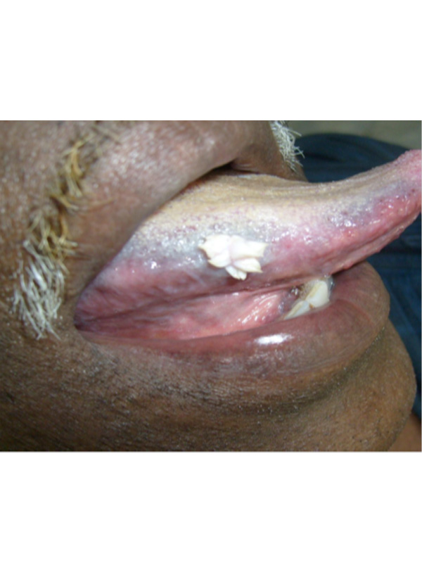
Oral squamous papilloma is a benign proliferation of stratified squamous epithelium, which results in a papillary or verrucous exophytic growth on the surface. Tomes in 1848 first described it as a localized wart on the gingiva which is an HPV-induced epithelial hyperplasia. It is usually reported in the age group of 30-50 years with no gender predilection, detected usually on the
buccal, labial or lingual mucosa. While most HPV lesions in other body parts are infective, oral papillomas are rarely contagious. It clinically displays a narrow stalk below a whitish growth bearing multiple blunted verruciform surface projections. The less keratinised lesions display a pinkish or reddish coloration. Submucosal fibrovascular connective tissues are contiguous with the stroma of the lesion. A small amount of scattered chronic inflammatory cells are common in the connective tissue stroma of the peduncle. The keratin on the surface is usually parakeratin and is thickened. Mild mitotic activity may be present rarely in this entity. Bearing striking clinical resemblance with verruca vulgaris, condyloma acuminatum or verruciform xanthoma, this condition can be histologically distinguished from them. Surgical excision is an adequate treatment for this condition and recurrence is rare. The figure displays the clinical features of a squamous papilloma in a 55 year old gentleman in whom the lesion was surgically removed.
 Awards Nomination