PHONE
+44-7482-878921
+44-7482-878921
2376-0249
Clinical-Medical Image - International Journal of Clinical & Medical Images (2023) Volume 10, Issue 7

Author(s): Mohamed AI Amine EI Mouden*
Department of Surgery, Abdelmalek Essaadi University, Tangier, Morocco
Received: 24 June 2023, Manuscript No. ijcmi-23-103711; Editor assigned: 26 June 2023, Pre QC No. P-103711; Reviewed: 14 July 2023, QC No. Q-103711; Revised: 18 July 2023, Manuscript No. R-103711; Published: 25 July 2023, DOI:10.4172/2376-0249.1000905
Citation: Mouden MAAE. (2023) Pelvic Hydatid Disease: Ultrasound is enough. Int J Clin Med Imaging 10: 905.
Copyright: © 2023 Mouden MAAE. This is an open-access article distributed under the terms of the Creative Commons Attribution License, which permits unrestricted use, distribution and reproduction in any medium, provided the original author and source are credited.
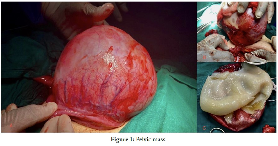
Hydatid cyst is a parasitic infection caused by Echinococcus granulosus, whose main host is the dog, and humans are considered accidental hosts. Morocco is an endemic country for hydatid disease. The liver is its most frequent location, followed by the lung. Pelvic hydatidosis is rare, with an incidence ranging from 0.30 to 4.27% of all hydatid cyst locations, according to different authors. Preoperative positive diagnosis is exceptional and is suspected in cases of pelvic symptoms associated with an abdominopelvic mass. Abdominopelvic ultrasound is sufficient for diagnosis in the majority of cases, while CT scan is useful in cases of diagnostic doubt and for differential diagnosis with other pelvic masses. The surgical treatment of choice is cystectomy. We report the case of a 40-year-old woman living in a rural area who presented with chronic pelvic pain for 3 years, without digestive or gynecological symptoms. Clinical examination revealed the presence of a well-defined, immobile 20 cm pelvic mass. Ultrasound confirmed the diagnosis of a stage III Gharbi lateral uterine hydatid cyst. Surgical exploration revealed a cystic formation adhering to the right tube and lateral face of the uterus, and a cystectomy was performed, preserving the right adnexa and taking care not to damage the ureter.
Pelvic hydatid disease; Parasitic infection; Cystectomy
None of the authors has any conflicts of interests to disclose.
 Awards Nomination