PHONE
+44-7482-878921
+44-7482-878921
2376-0249
Case Blog - International Journal of Clinical & Medical Images (2018) Volume 5, Issue 12

Author(s): Arregger Alejandro Luis*, Cardoso Estela Maria del Lujan and Contreras Liliana Noemi
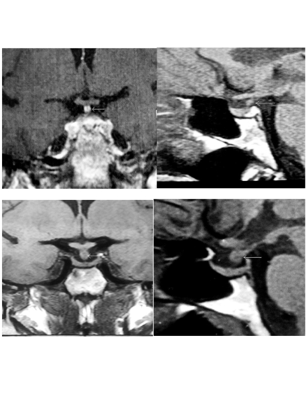
Patient 1: A 64 year old woman was admitted to the emergency room for vomiting and high blood pressure. Otherwise normal, she had been suffering from proximal muscle weakness, fatigue, reduced appetite, cognitive impairment and pre-tibial myxedema during the last two months. The patient denied glucocorticoid intake. Mild hyponatremia (132 mEq/l) and anemia (hct: 30%) were detected. TSH was high (13.74 µU/ml) with low FT4: 0.57ng/dl. Anti-peroxidase antibodies were positive. Serum potassium was normal (4.2 mEq/l). Serum morning cortisol was less than 1.0 µg/dl and ACTH was 6.7 pg/ml and 5.0 pg/ ml in two different occasions. An ACTH stimulating test (Synacthen 250.0 µg, i.m) showed a blunted cortisol response at 30 and 60 min after stimulus, confirming adrenal insufficiency. The remaining pituitary function was normal. MRI showed a thickened pituitary stalk (Figure 1). The patient improved on hydrocortisone and LT4.
Patient 2: A 30 year old woman complained of amenorrhea and polyuria (9 L/day). She had high plasma and low urinary osmolality (311 mOsm/kg and 136 mOsm/kg, respectively). Low serum estradiol and gonadotropins were found. Thyroid and adrenal functions were normal. MRI showed a thickened pituitary stalk (Figure 2). She started on desmopressin (0.1 mg/day) which reduced daily diuresis by 60% and lowered plasma osmolality (290 mOsm/day). Additionally she began on estrogen and progestin replacement.
Discussion: Pituitary stalk lesions may be found by chance or during the evaluation of endocrine disorders. In a series of 152 patients, the etiologies were attributed to neoplasia in 32%, inflammatory lesions 20%, and congenital anomalies in 9% and in 39% remained unknown [2]. Primary hypophysitis is rare, between 0.2 and 6.5% among pituitary pathologies. Three histopathological types are described in this category: lymphocytic, granulomatous and xanthomatous hypophysitis [3].These conditions are usually confined to the pituitary. In secondary hypophysitis, a systemic disease is the cause of the pituitary lesion: Sarcoidosis, Wegener granulomatosis, Langerhans cell histyocitosis and IgG4 related plasmatic histiocytosis may affect the pituitary. Monoclonal antibodies against the cytotoxic T-lymphocyte antigen-4 (CTLA-4) for treating metastatic cancer, may induce hypophysitis [4,5]. In the setting of immunosuppression, fungal infection, tuberculosis and toxoplasmosis may involve the pituitary gland [4]. Interestingly, these two patients showed different clinical presentations. After excluding secondary etiologies, primary hypophysitis was suspected in both [4]. Although histological confirmation is needed, biopsy of a pituitary stalk lesion is technically demanding, and must be performed by an experienced neurosurgeon. In order to avoid invasive procedures, the approach was based on clinical, biochemical and radiological features. A close follow-up is mandatory to detect new symptoms and to determine if the lesions progress over time.
 Awards Nomination