PHONE
+44-7482-878921
+44-7482-878921
2376-0249
Clinical-Medical Image - International Journal of Clinical & Medical Images (2023) Volume 10, Issue 5

Author(s): Fatima Zohra Benbrahim*, Asaad EL Bakkari , Hajar Zebbakh, Hatim Essaber, Soukaina Allioui, Hounayda Jerguigue, Youssef Omor and Rachida Latib
Department of Radiology, National Institute of Oncology, UHC Ibn Sina, Mohamed V University, Rabat, Morocco
Received: 12 April 2023, Manuscript No. ijcmi-23-95227; Editor assigned: 14 April 2023, Pre QC No. P-95227; Reviewed: 28 April 2023, QC No. Q-95227; Revised: 03 May 2023, Manuscript No. R-95227; Published: 10 May 2023, DOI:10.4172/2376-0249.1000893
Citation: Benbrahim FZ, Bakkari AEI, Zebbakh H, Essaber H and Allioui S, et al. (2023) Poke Ball Sign: Mature Cystic Teratoma. Int J Clin Med Imaging 10: 893.
Copyright: © 2023 Benbrahim FZ, et al. This is an open-access article distributed under the terms of the Creative Commons Attribution License, which permits unrestricted use, distribution, and reproduction in any medium, provided the original author and source are credited.
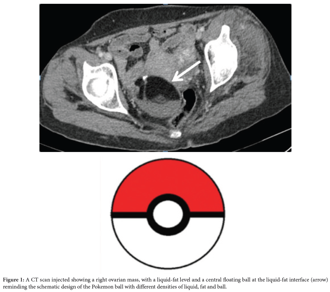
A 38-year-old woman, genitally active, recently diagnosed with breast cancer, in whom a thoraco-abdomino-pelvic CT scan with contrast injection performed as part of the extension workup revealed a right ovarian mass with a fat-liquid level and a round central nodule floating at the fat-liquid interface, giving the appearance of a poke ball (Figure 1).
Poke ball sign is a pathognomonic radiological sign of mature cystic teratoma, characterized by the presence of a single flotting ball at the liquidfat interface. Poke Ball Sign is a variant of «Flotting ball sign» which corresponds to the presence of a variable number of mobile balls within the cystic and fatty contents of an adnexal mass [1]. Mature cystic teratoma is the most frequent benign ovarian tumor in reproductive age women. It is often asymptomatic and discovered incidentally on imaging [2] or more rarely with pelvic pain or complications such as rupture, adnexal torsion or malignant degeneration. To various degrees, these floating balls are constituted by debris of sebum, keratin, fibrin, hemosiderin or fat [3], which influence their disposition in the cystic area and their appearance on imaging. The identification of a floating ball at the fluid-fat interface is often easier on CT and on MRI imaging [4]. Mature teratoma takes on different aspects on imaging, although the poke ball sign is pathognomonic, it has a low sensitivity (25 to 30% in some case series) which explains its absence doesn’t formally eliminate the diagnosis of mature teratoma [1,4].
Poke ball sign; Teratoma; Ovary
The authors declare that they have no conflict of interest in relation to this article.
[1] Şahin H, Akdoğan AI, Ayaz D, Karadeniz T and Sancı M. (2019). Utility of the “floating ball sign” in diagnosis of ovarian cystic teratoma. Turk J Obstet Gynecol 16: 118.
Google Scholar, Crossref, Indexed at
[2] Maslin P, Luchs JS, Haas J and Katz DS. (2002). Ovarian teratoma with malignant transformation: CT diagnosis. AJR Am J Roentgenol 178: 1574-1574.
Google Scholar, Crossref, Indexed at
[3] Choudhary S, Fasih N, Innes MM and Marginean C. (2009). Imaging of ovarian teratomas: Appearances and complications. J Med Imaging Radiat Oncol 53: 480-488.
Google Scholar, Crossref, Indexed at
[4] Kim HC, Kim SH, Lee HJ, Shin SJ and Hwang SI, et al. (2002). Fluid–fluid levels in ovarian teratomas. Abdom Imaging 27, 100-105.
 Awards Nomination