PHONE
+44-7482-878921
+44-7482-878921
2376-0249
Clinical-Medical Image - International Journal of Clinical & Medical Images (2020) Volume 7, Issue 11

Author(s): Olaia Chalh*, Jerguigue H, Latib R and Omor Y
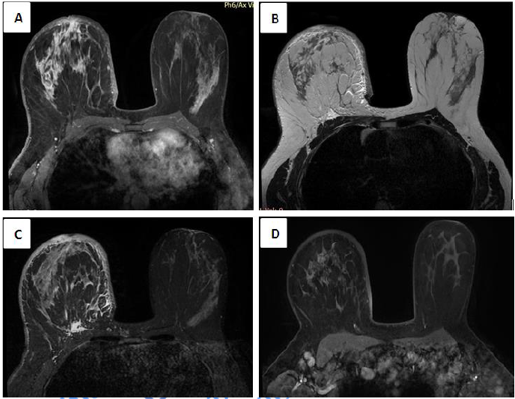
Magnetic Resonance Mammography (MRM) is the most accurate imaging modality in the detection of breast cancer. On the basis of morphologic and kinetic sequences (especially T2-weighted), it may differentiate between benign and malignant lesions, and contributes to the final diagnosis. Some MRI findings have a diagnostic and prognostic value. Defined as high SI on T2WI in the retromammary area, PE is a specific MRI finding of inflammatory breast cancer (IBC) and its occult forms (Figure 1). Besides its low prevalence (9%), PE is considered as a high significant indicator for other malignant tumors with a high specificity (97.3%) and positive predictive value (100%). It is often associated with invasive carcinomas >2 cm in diameter, independent from histological types. Prepectoral edema results from infiltrated lymphovascular system by tumor cells. Breast cancer often affects axillary lymph node leading to an obstruction of lymphatic trails. Drainage to the internal mammary and interpectoral nodes could be the main lymphatic drainage route. After being involved with cancer cells, perepectoral edema appears. In MR-mammography, the presence of prepectoral edema is indicative of advanced stage tumors with extensive lymphovascular invasion and high axillary lymph node positivity. It might predict poor prognosis patients, requiring more intense treatment.
 Awards Nomination