PHONE
+44-7482-878921
+44-7482-878921
2376-0249
Medical Image - International Journal of Clinical & Medical Images (2015) Volume 2, Issue 4

Author(s): Alawad AAM* and Gismalla MD
A 20-year-old female presented with a complaint of a progressive swelling in the left lower abdomen for the last three months. There was no history of preceding trauma, fever, cough, malaise or pain. There was no history of contact with any case of tuberculosis. On examination, there was swelling in the left iliac fossa measuring 8 × 8 cm in size, non-tender with smooth and ill-defined margins and a normal overlying skin. Examinations of the cardiovascular and respiratory system were within normal limits. Laboratory investigations were within normal limits. ELISA for HIV was negative. The chest radiograph was unremarkable.
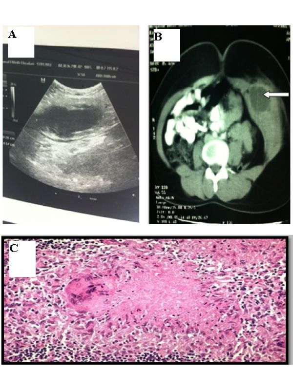
Abdominal ultrasonography of the abdomen revealed a 6.5 × 8.5 cm left iliac fossa cystic mass with a liquefied necrotic center in the anterior abdominal wall (Figure 1A). Computerized Tomography scan of the abdomen showed an abscess in the left antero-lateral portion of the abdominal wall limited to the muscle layer (Figure 1B). Ultrasound-guided fine-needle aspiration and cytological examination revealed caseating granuloma with central necrosis, lymphocytes, and giant cells, consistent with tuberculosis (Figure 1C). The patient was diagnosed to have tuberculous abscess of the anterior abdominal wall and antituberculosis treatment was started. After four weeks’ antituberculous treatment, she responded well to the treatment and the abscess regressed considerably. The prognosis is good in tuberculous myositis with appropriate chemotherapy [1,2]. This case cautions the clinicians and radiologists about the possibility of tuberculosis in considering the differential diagnosis of any lesion even in any unlikely anatomical area, especially in those areas where tuberculosis is endemic.
 Awards Nomination