PHONE
+44-7482-878921
+44-7482-878921
2376-0249
Clinical-Medical Image - International Journal of Clinical & Medical Images (2023) Volume 10, Issue 1

Author(s): Dan Ramsey*
Department of Exercise Science, School of Public Health and Health Professions, University at Buffalo, Buffalo, NY 14260, USA
Received: 03 January 2023, Manuscript No. ijcmi-23-96507; Editor assigned: 04 January 2023, Pre QC No. P-96507; Reviewed: 18 January 2023, QC No. Q-96507; Revised: 23 January 2023, Manuscript No. R-96507; Published: 30 January 2023, DOI:10.4172/2376-0249.1000874
Citation: Ramsey D. (2023) Radiology’s Role in Diagnosing and Treating Dislocated Elbow Injuries. Int J Clin Med Imaging 10:874.
Copyright: © 2023 Ramsey D. This is an open-access article distributed under the terms of the Creative Commons Attribution License, which permits unrestricted use, distribution, and reproduction in any medium, provided the original author and source are credited.
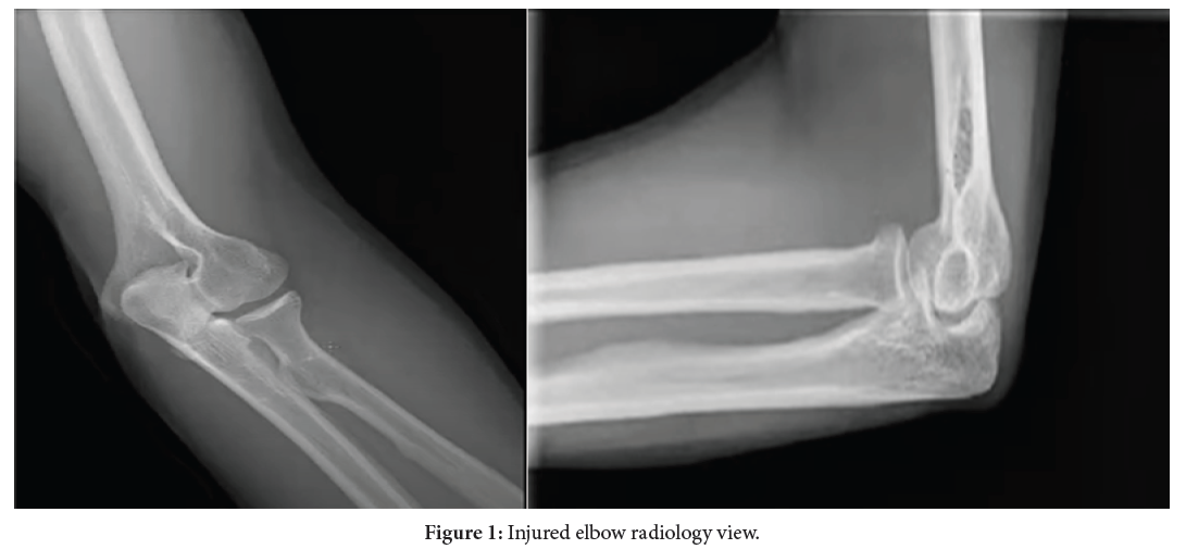
A dislocated elbow occurs when the bones that make up the elbow joint are forced out of their normal position. Radiology is a medical specialty that uses imaging techniques such as X-rays, CT scans and MRI to diagnose and treat medical conditions. In the case of a dislocated elbow, radiology plays an important role in confirming the diagnosis and determining the severity of the injury. X-rays are typically the first imaging test performed to assess the extent of the dislocation and to rule out any associated fractures. On an X-ray, a dislocated elbow may appear as a gap between the bones that make up the joint or a visible displacement of the bones. CT scans and MRI may also be used to obtain more detailed images of the elbow joint and surrounding structures, which can be useful in planning for surgical intervention if necessary. It’s important to seek medical attention promptly if you suspect that you have dislocated your elbow. Treatment may involve resetting the joint and immobilizing the arm in a cast or brace to allow for healing. In some cases, surgery may be necessary to repair any damaged structures or to realign the joint [1,2].
Nerve reconstruction; Radiology; Brachial plexus injury; Elbow injuries
None.
The authors declare no conflict of interest.
[1] Leechavengvongs S, Witoonchart K, Uerpairojkit C, Thuvasethakul P and Malungpaishrope K. (2006). Combined nerve transfers for C5 and C6 brachial plexus avulsion injury. Hand Surg 31: 183-189.
Google Scholar, Crossref, Indexed at
[2] Bertelli JA and Ghizoni MF. (2004). Reconstruction of C5 and C6 brachial plexus avulsion injury by multiple nerve transfers: Spinal accessory to suprascapular, ulnar fascicles to biceps branch and triceps long or lateral head branch to axillary nerve. Hand Surg 29: 131-139.
 Awards Nomination